E.coli-derived human Galectin 3 recombinant protein (Position: K139-I250). Human Galectin 3 shares 88% and 84% amino acid (aa) sequence identity with mouse and rat Galectin 3, respectively.
Konjugation:
Unconjugated
Alternative Synonym:
35 kDa lectin, CBP 35, CBP35, Gal 3, GAL3, Galactose specific lectin 3, Galactoside binding protein, GALBP, Galectin 3, Galectin-3, GALIG, IgE binding protein, L 31, Laminin binding protein, Lectin L 29, LGALS2, LGALS3, Mac 2 antigen, MAC2
Anti-Galectin 3/LGALS3 Antibody (monoclonal, 12B12). Tested in Flow Cytometry, IF, IHC, ICC, WB applications. This antibody reacts with Human, Mouse, Rat.
Klonalität:
Monoclonal
Konzentration:
Adding 0.2 ml of distilled water will yield a concentration of 500 µg/ml.
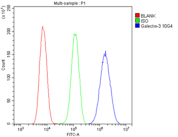
Flow Cytometry analysis of U20S cells using anti-Galectin 3/LGALS3 antibody. Overlay histogram showing U20S cells (Blue line). To facilitate intracellular staining, cells were fixed with 4% paraformaldehyde and permeabilized with permeabilization buffer. The cells were blocked with 10% normal goat serum. And then incubated with mouse anti-Galectin 3/LGALS3 Antibody (1 µg/1x10 6 cells) for 30 min at 20C. DyLight488 conjugated goat anti-mouse IgG (5-10 µg/1x10 6 cells) was used as secondary antibody for 30 minutes at 20C. Isotype control antibody (Green line) was mouse IgG (1 µg/1x10 6) used under the same conditions. Unlabelled sample (Red line) was also used as a control.
WB analysis using anti-Galectin 3/LGALS3 antibody.Lane 1
IF analysis of Galectin 3/LGALS3 using anti-Galectin 3/LGALS3 antibody. Galectin 3/LGALS3 was detected in immunocytochemical section of A431 cells. Enzyme antigen retrieval was performed using IHC enzyme antigen retrieval reagent for 15 mins. The cells were blocked with 10% goat serum. And then incubated with 2 µg/mL mouse anti-Galectin 3/LGALS3 Antibody overnight at 4C. DyLight488 Conjugated Goat Anti-Mouse IgG was used as secondary antibody at 1:100 dilution and incubated for 30 minutes at 37C. The section was counterstained with DAPI. Visualize using a fluorescence microscope and filter sets appropriate for the label used.
IF analysis of Galectin 3/LGALS3 using anti-Galectin 3/LGALS3 antibody. Galectin 3/LGALS3 was detected in immunocytochemical section of MCF-7 cells. Enzyme antigen retrieval was performed using IHC enzyme antigen retrieval reagent for 15 mins. The cells were blocked with 10% goat serum. And then incubated with 2 µg/mL mouse anti-Galectin 3/LGALS3 Antibody overnight at 4C. DyLight488 Conjugated Goat Anti-Mouse IgG was used as secondary antibody at 1:100 dilution and incubated for 30 minutes at 37C. The section was counterstained with DAPI. Visualize using a fluorescence microscope and filter sets appropriate for the label used.
IHC analysis of Galectin 3/LGALS3 using anti Galectin 3/LGALS3 antibody. Galectin 3/LGALS3 was detected in paraffin-embedded section of human intestinal cancer tissue. Heat mediated antigen retrieval was performed in EDTA buffer (pH8.0, epitope retrieval solution). The tissue section was blocked with 10% goat serum. The tissue section was then incubated with 1 µg/ml mouse anti-Galectin 3/LGALS3 Antibody overnight at 4C. Biotinylated goat anti-mouse IgG was used as secondary antibody and incubated for 30 minutes at 37C. The tissue section was developed using Strepavidin-Biotin-Complex (SABC) with DAB as the chromogen.
IHC analysis of Galectin 3/LGALS3 using anti Galectin 3/LGALS3 antibody. Galectin 3/LGALS3 was detected in paraffin-embedded section of human melanoma tissue. Heat mediated antigen retrieval was performed in EDTA buffer (pH8.0, epitope retrieval solution). The tissue section was blocked with 10% goat serum. The tissue section was then incubated with 1 µg/ml mouse anti-Galectin 3/LGALS3 Antibody overnight at 4C. Biotinylated goat anti-mouse IgG was used as secondary antibody and incubated for 30 minutes at 37C. The tissue section was developed using Strepavidin-Biotin-Complex (SABC) with DAB as the chromogen.
IHC analysis of Galectin 3/LGALS3 using anti Galectin 3/LGALS3 antibody. Galectin 3/LGALS3 was detected in paraffin-embedded section of mouse small intestine tissue. Heat mediated antigen retrieval was performed in EDTA buffer (pH8.0, epitope retrieval solution). The tissue section was blocked with 10% goat serum. The tissue section was then incubated with 1 µg/ml mouse anti-Galectin 3/LGALS3 Antibody overnight at 4C. Biotinylated goat anti-mouse IgG was used as secondary antibody and incubated for 30 minutes at 37C. The tissue section was developed using Strepavidin-Biotin-Complex (SABC) with DAB as the chromogen.
IHC analysis of Galectin 3/LGALS3 using anti Galectin 3/LGALS3 antibody. Galectin 3/LGALS3 was detected in paraffin-embedded section of rat small intestine tissue. Heat m
* Mehrwertsteuer und Versandkosten nicht enthalten. Irrtümer und Preisänderungen vorbehalten