Application Notes: Western blot, 0.1-0.5µg/ml Immunohistochemistry (Paraffin-embedded Section), 0.5-1µg/ml Immunocytochemistry/Immunofluorescence, 2µg/ml Flow Cytometry (Fixed), 1-3µg/1x106 cells ELISA (Cap), 1-5µg/ml. Add 0.2ml of distilled water will yield a concentration of 500µg/ml
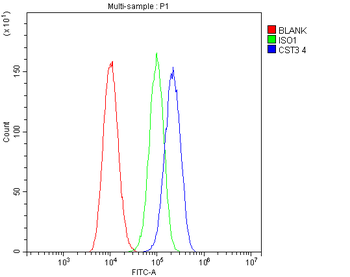
Flow Cytometry analysis of A549 cells using anti-Cystatin C/CST3 antibody. Overlay histogram showing A549 cells (Blue line). To facilitate intracellular staining, cells were fixed with 4% paraformaldehyde and permeabilized with permeabilization buffer. The cells were blocked with 10% normal goat serum. And then incubated with mouse anti-Cystatin C/CST3 Antibody (1 µg/1x10 6 cells) for 30 min at 20C. DyLight488 conjugated goat anti-mouse IgG (5-10 µg/1x10 6 cells) was used as secondary antibody for 30 minutes at 20C. Isotype control antibody (Green line) was mouse IgG (1 µg/1x10 6) used under the same conditions. Unlabelled sample (Red line) was also used as a control.
WB analysis of Cystatin C/CST3 using anti-Cystatin C/CST3
IF analysis of CST3 using anti-CST3 antibody. CST3 was detected in immunocytochemical section of MCF7 cells. Enzyme antigen retrieval was performed using IHC enzyme antigen retrieval reagent for 15 mins. The cells were blocked with 10% goat serum. And then incubated with 2 µg/mL mouse anti-CST3 Antibody overnight at 4C. DyLight488 Conjugated Goat Anti-Mouse IgG was used as secondary antibody at 1:100 dilution and incubated for 30 minutes at 37C. The section was counterstained with DAPI. Visualize using a fluorescence microscope and filter sets appropriate for the label used.
IHC analysis of Cystatin C/CST3 using anti-Cystatin C/CST3 antibody. Cystatin C/CST3 was detected in paraffin-embedded section of human colon cancer tissue. Heat mediated antigen retrieval was performed in EDTA buffer (pH8.0, epitope retrieval solution). The tissue section was blocked with 10% goat serum. The tissue section was then incubated with 1 µg/ml mouse anti-Cystatin C/CST3 Antibody overnight at 4C. Biotinylated goat anti-mouse IgG was used as secondary antibody and incubated for 30 minutes at 37C. The tissue section was developed using Strepavidin-Biotin-Complex (SABC) with DAB as the chromogen.
IHC analysis of Cystatin C/CST3 using anti-Cystatin C/CST3 antibody. Cystatin C/CST3 was detected in paraffin-embedded section of human colon cancer tissue. Heat mediated antigen retrieval was performed in EDTA buffer (pH8.0, epitope retrieval solution). The tissue section was blocked with 10% goat serum. The tissue section was then incubated with 1 µg/ml mouse anti-Cystatin C/CST3 Antibody overnight at 4C. Biotinylated goat anti-mouse IgG was used as secondary antibody and incubated for 30 minutes at 37C. The tissue section was developed using Strepavidin-Biotin-Complex (SABC) with DAB as the chromogen.
IHC analysis of Cystatin C/CST3 using anti-Cystatin C/CST3 antibody. Cystatin C/CST3 was detected in paraffin-embedded section of human glioma tissue. Heat mediated antigen retrieval was performed in EDTA buffer (pH8.0, epitope retrieval solution). The tissue section was blocked with 10% goat serum. The tissue section was then incubated with 1 µg/ml mouse anti-Cystatin C/CST3 Antibody overnight at 4C. Biotinylated goat anti-mouse IgG was used as secondary antibody and incubated for 30 minutes at 37C. The tissue section was developed using Strepavidin-Biotin-Complex (SABC) with DAB as the chromogen.
Sandwich ELISA - Recombinant human Cystatin C/CST3 protein standard curve. Use in combination with reagents from Human Cystatin C/CST3 ELISA Kit EZ-Set (DIY Antibody Pairs).
Western blot analysis of Cystatin C/CST3 using anti-Cystatin C/CST3 antibody. Electrophoresis was performed on a 5-20% SDS-PAGE gel at 70V (Stacking gel) / 90V (Resolving gel) for 2-3 hours. The sample well of each lane was loaded with 50 ug of sample under reducing conditions. Lane 1: human PC-3 whole cell lysates, Lane 2: human CACO-2 whole cell lysates, Lane 3: human placenta tissue lysates, Lane 4: human A549 whole cell lysates, Lane 5: human COLO320 whole cell lysates, Lane 6: human HEK293 whole cell lysates. After Electrophoresis, proteins were transferred to a Nitrocellulose membrane at 150mA for 50-90 minutes. Blocked the membrane with 5% Non-fat Milk/ TBS for 1.5 hour at RT. The membrane was incubated with
* Mehrwertsteuer und Versandkosten nicht enthalten. Irrtümer und Preisänderungen vorbehalten