The immunogen is a Red Fluorescent Protein (RFP) fusion protein corresponding to the full-length amino acid sequence (234aa) derived from the mushroom anemone Discosoma.
Konjugation:
Unconjugated
Alternative Synonym:
DsRed protein, rDsRed, Discosoma sp. Red Fluorescent Protein, Red fluorescent protein drFP583, sea anemone Discosoma sp. Mushroom, RFP antibody, RFP-RTU, Ready To Use Antibody, RTU Antibody
RTU Anti-RFP was prepared from monospecific antiserum by immunoaffinity chromatography using Red Fluorescent Protein (Discosoma) coupled to agarose beads followed by solid phase adsorption(s) to remove any unwanted reactivities. Expect reactivity against RFP and its variants: mCherry, tdTomato, mBanana, mOrange, mPlum, mOrange and mStrawberry. Assay by immunoelectrophoresis resulted in a single precipitin arc against anti-Rabbit Serum and purified and partially purified Red Fluorescent Protein (Discosoma). No reaction was observed against Human, Mouse or Rat serum proteins.
Formulierung:
Liquid (sterile filtered)
Application Verdünnung:
WB: 1:1,000
Anwendungsbeschreibung:
Application Notes: Ready-To-Use Anti-RFP is designed to detect RFP and its variants. Ready-To-Use Anti-RFP Rabbit Polyclonal Antibody has been optimized and tested in ELISA and in western blot using 1:1000 dilution. This Anti-RFP (RTU) Antibody is sufficient to run 10 western blots. Although not tested, this antibody is likely functional in immunohistochemistry, immunofluorescence, and immunoprecipitation. Optimal titers for these applications should be determined by the researcher
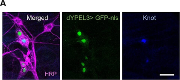
dYPEL3 frameshift mutations reduce nociceptive behavior. (A) Nociceptive/class IV da neurons are positive for dYPEL3. A nuclear GFP (GFP-nls, green) was expressed under dYPEL3-GAL4 following immunostaining with anti-Knot antibody (blue). Anti-HRP antibody was used to label all PNS neurons (magenta). Scale bar: 10 µm. (B) The AITC-induced nociceptive behavior was measured in a wild-type control (wt) and dYPEL3 frameshift mutants (dYPEL3T1-8 and dYPEL3T1-6). The number of larvae that exhibited complete rolling behavior was scored and expressed as a percentage (n = 252 for each genotype). The Chi-squared test was performed between the groups. NS, non-significant, ****P < 0.0001.
dYPEL3 frameshift mutations reduce the synaptic contact between nociceptors and Basin-4 neurons. (A) The syb-GRASP technique was used to report the synaptic contact between nociceptors and Basin-4. The spGFP1-10 (red cylinders) and spGFP11 (blue sectors) were expressed in nociceptors and Basin-4, respectively (left). The resulting GRASP signal was visualized by anti-GRASP antibody (green), and the spGFP1-10 that is expressed in nociceptor axon terminals was used as a normalization control (magenta) (right). Scale bar: 10 µm. (B) The GRASP intensity from each neuropil was normalized by spGFP1-10 intensity and presented as means.e.m. (left), as well as in a violin plot to show distribution (right) (n = 36 for wt, n = 34 for dYPEL3T1-8). Mann-Whitney test. All statistical analysis was two-tailed. **P < 0.01.
dYPEL3 is a neuronal gene. (A) The expression pattern of dYPEL3 in the CNS. The InSITE gene trap line for dYPEL3 was used (CG15309-GAL4/dYPEL3-GAL4). GAL4 transcription factor is inserted in the first intron. The introduction of UAS-mCD8::GFP demonstrates the endogenous expression pattern of dYPEL3. Note that the CG15309-GAL-positive cells elaborate fine processes throughout the CNS. Scale bar: 50 µm. (B) mCD8::GFP (magenta) was expressed under dYPEL3-GAL4 following immunostaining with anti-Elav (neuronal, green) and anti-Repo (glial, blue) antibodies. (i) The CNS. Cell bodies in 1 and 2 are shown magnified on the top right. (ii, iii) Chordotonal neurons (ii, arrows) and a class III da neuron (iii) in the PNS. iii also shows a class IV da neuron (nociceptor) that is positive for dYPEL3 (arrow). Scale bar: 10 µm. (C) Quantitation of the Elav-positive and Repo-positive cells that are labeled with CG15309-GAL4. The majority of dYPEL3-postive cells were Elav positive, but none were positive for Repo.
dYPEL3 knockout mutants recapitulate the phenotypes in the dYPEL3 frameshift mutants. (A) The generation of dYPEL3 knockout (dYPEL3KO) flies. Top: the entire dYPEL3 ORF was deleted using CRISPR/Cas9-mediated homology-directed recombination. Bottom: agarose gel image of PCR-based genotyping. Note that dYPEL3KO showed the PCR amplifications specific for DsRed-cassette, but not the ones from wild type. (B) dYPEL3 knockout reduces AITC-induced behavioral responses. AITC-induced nociceptive behavior was measured in a wild-type control (wt) and dYPEL3 knockout mutants (dYPEL3KO). The number of larvae that exhibited complete rolling behavior was scored and expressed as a percentage (n = 252 for wt, n = 203 for dYPEL3KO). The Chi-squared test was performed between the groups. (C) dYPEL3 knockout caused a reduction in Basin-4 activation upon AITC. GCaMP6f was expressed in Basin-4 neurons. Nociceptors were activated with 10 mM AITC. Ca2 + increase in Basin-4 was measured by GCaMP fluorescence and the GCaMP trace over time is shown (n = 47 for wt, n = 46 for dYPEL3KO) (left). The cumulative GCaMP activation from single Basin-4 neurons was measured and presented as means.e.m. (middle), as well as in a violin plot to show distribution (right). Mann-Whitney test. (D) dYPEL3 knockout causes a reduction in syb-GRASP signals between nociceptors and Basin-4. Synaptobrevin-spGFP1-10 and spGFP11 were expressed in nociceptors and Basin-4, respectively. The resulting GRASP signal
* Mehrwertsteuer und Versandkosten nicht enthalten. Irrtümer und Preisänderungen vorbehalten