136.36mM Ethanolamine, 133.23 mM Chlorides, 9.55mM Phosphates, 9.55mM Sodium Bicarbonate
Target-Kategorie:
HSP70
Application Verdünnung:
WB (1:1000), ICC/IF (1:100), FACS (1:250)
Anwendungsbeschreibung:
Application Notes: A 1:250 dilution of SMC-249 was sufficient for detection of cell surface HSP70 in HCT116 cells using Fluorescence-activated cell sorting, applied in flow cytometry, with FITC as the fluorescent probe
Fluorescence-activated cell sorting analysis using Mouse Anti-HSP70 Monoclonal Antibody, Clone 1H11. Tissue: Jurkat E6.1 cells. Species: Human. Fixation: No fixation. Primary Antibody: Mouse Anti-HSP70 Monoclonal Antibody at 20 µg/ml for 40 min at 4C. Counterstain: Propidium Iodide nuclear stain at 2.5 µg/ml for 5 min at RT. Isotype Control: Anti-mouse FITC at 1:32 for 15 min at RT (blue line).
Western Blot analysis of Human Heat Shocked cervical cancer cell line (HeLa) lysate showing detection of HSP70 protein using Mouse Anti-HSP70 Monoclonal Antibody, Clone 1H11. Lane 1: Molecular Weight ladder (MW). Lane 2: HeLa cell lysates. Load: 20 µg. Primary Antibody: Mouse Anti-HSP70 Monoclonal Antibody at 1:1000.
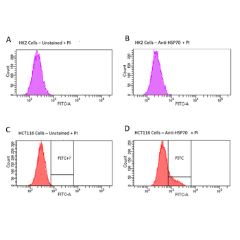
Fluorescence-activated cell sorting analysis using Mouse Anti-HSP70 Monoclonal Antibody, Clone 1H11. Tissue: HCT116 and HK2 cells. Species: Human. Primary Antibody: Mouse Anti-HSP70 Monoclonal Antibody at 1:100 for 90 min at 4C. Counterstain: Propidium Iodide nuclear stain. (A) HK2 cells unstained. (B) HK2 cells and HSP70 Antibody. (C) HCT116 cells unstained. (D) HCT116 cells and HSP70 Antibody. Shows that HSP70 Antibody binds to the cell surface of tumor cells but not non-tumor cells.
Fluorescence-activated cell sorting analysis using Mouse Anti-HSP70 Monoclonal Antibody, Clone 1H11. Tissue: Jurkat E6.1, U937, MCF7 and HT29 cells. Species: Human. Fixation: No fixation. Primary Antibody: Mouse Anti-HSP70 Monoclonal Antibody at 20 µg/ml for 40 min at 4C. Counterstain: Propidium Iodide nuclear stain at 2.5 µg/ml for 5 min at 4C. Shows that binding of HSP70 Antibody is not cell-line specific.