ATP citrate lyase ACLY Mouse Monoclonal Antibody, Clone: [5I2], Unconjugated
Artikelnummer:
BYT-ORB548019
Hersteller Artikelnummer:
orb548019
Alternativnummer:
BYT-ORB548019-100
Hersteller:
Biorbyt
Wirt:
Mouse
Kategorie:
Antikörper
Applikation:
FC, ICC, IF, IHC, WB
Spezies Reaktivität:
Human, Mouse, Rat
Immunogen:
E. coli-derived human ATP citrate lyase recombinant protein (Position: M1-I180). Human ATP citrate lyase shares 95% amino acid (aa) sequence identity with both mouse and rat ATP citrate lyase.
Konjugation:
Unconjugated
Alternative Synonym:
ACL, ACLY, ATP citrate (pro S ) lyase, ATP citrate lyase, ATP citrate synthase, ATPCL, Citrate cleavage enzyme, CLATP
Anti-ATP citrate lyase ACLY Antibody (monoclonal, 5I2). Tested in Flow Cytometry, IF, IHC, ICC, WB applications. This antibody reacts with Human, Mouse, Rat.
Western blot, 0.1-0.5µg/ml Immunohistochemistry (Paraffin-embedded Section), 0.5-1µg/ml Immunocytochemistry/Immunofluorescence, 2µg/ml, Human Flow Cytometry (Fixed), 1-3µg/1x10 6 cells
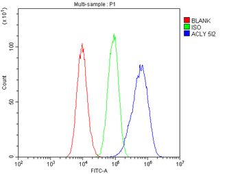
Flow Cytometry analysis of A549 cells using anti-ATP citrate lyase antibody. Overlay histogram showing A549 cells (Blue line). To facilitate intracellular staining, cells were fixed with 4% paraformaldehyde and permeabilized with permeabilization buffer. The cells were blocked with 10% normal goat serum. And then incubated with mouse anti-ATP citrate lyase Antibody (1 µg/1x10 6 cells) for 30 min at 20C. DyLight488 conjugated goat anti-mouse IgG (5-10 µg/1x10 6 cells) was used as secondary antibody for 30 minutes at 20C. Isotype control antibody (Green line) was mouse IgG (1 µg/1x10 6) used under the same conditions. Unlabelled sample (Red line) was also used as a control.
IF analysis of ATP citrate lyase using anti-ATP citrate lyase antibody. ATP citrate lyase was detected in immunocytochemical section of MCF7 cells. Enzyme antigen retrieval was performed using IHC enzyme antigen retrieval reagent for 15 mins. The cells were blocked with 10% goat serum. And then incubated with 2 µg/mL mouse anti-ATP citrate lyase Antibody overnight at 4C. DyLight488 Conjugated Goat Anti-Mouse IgG was used as secondary antibody at 1:100 dilution and incubated for 30 minutes at 37C. The section was counterstained with DAPI. Visualize using a fluorescence microscope and filter sets appropriate for the label used.
IHC analysis of ATP citrate lyase using anti-ATP citrate lyase antibody. ATP citrate lyase was detected in paraffin-embedded section of human pancreatic cancer tissue. Heat mediated antigen retrieval was performed in EDTA buffer (pH8.0, epitope retrieval solution). The tissue section was blocked with 10% goat serum. The tissue section was then incubated with 1 µg/ml mouse anti-ATP citrate lyase Antibody overnight at 4C. Biotinylated goat anti-mouse IgG was used as secondary antibody and incubated for 30 minutes at 37C. The tissue section was developed using Strepavidin-Biotin-Complex (SABC) with DAB as the chromogen.
IHC analysis of ATP citrate lyase using anti-ATP citrate lyase antibody. ATP citrate lyase was detected in paraffin-embedded section of human testis cancer tissue. Heat mediated antigen retrieval was performed in EDTA buffer (pH8.0, epitope retrieval solution). The tissue section was blocked with 10% goat serum. The tissue section was then incubated with 1 µg/ml mouse anti-ATP citrate lyase Antibody overnight at 4C. Biotinylated goat anti-mouse IgG was used as secondary antibody and incubated for 30 minutes at 37C. The tissue section was developed using Strepavidin-Biotin-Complex (SABC) with DAB as the chromogen.
IHC analysis of ATP citrate lyase using anti-ATP citrate lyase antibody. ATP citrate lyase was detected in paraffin-embedded section of mouse pancreas tissue. Heat mediated antigen retrieval was performed in EDTA buffer (pH8.0, epitope retrieval solution). The tissue section was blocked with 10% goat serum. The tissue section was then incubated with 1 µg/ml mouse anti-ATP citrate lyase Antibody overnight at 4C. Biotinylated goat anti-mouse IgG was used as secondary antibody and incubated for 30 minutes at 37C. The tissue section was developed using Strepavidin-Biotin-Complex (SABC) with DAB as the chromogen.
IHC analysis of ATP citrate lyase using anti-ATP citrate lyase antibody. ATP citrate lyase was detected in paraffin-embedded section of rat pancreas tissue. Heat mediated antigen retrieval was performed in EDTA buffer (pH8.0, epitope retrieval solution). The tissue section was blocked with 10% goat serum. The tissue section was then incubated with 1 µg/ml mouse anti-ATP citrate lyase Antibody overnight at 4C. Biotinylated goat anti-mouse IgG was used as secondary antibody and incubated for 30 minutes at 37C. The tissue section was developed using Strepavidin-Biotin-Complex (SABC) with DAB as the chromogen.
Western blot analysis of ATP citrate lyase using anti-ATP citrate lyase antibody. Electrophoresis was performed on a 5-20% SDS-PAGE gel at 70V (Stacking gel) / 90V (Resolving gel) for 2-3 hours.
* Mehrwertsteuer und Versandkosten nicht enthalten. Irrtümer und Preisänderungen vorbehalten