
Cytokeratin 1 Recombinant Rabbit Monoclonal Antibody, Clone: [1C2], Unconjugated
Artikelnummer:
BYT-ORB559083
- Bilder (9)


| Artikelname: | Cytokeratin 1 Recombinant Rabbit Monoclonal Antibody, Clone: [1C2], Unconjugated |
| Artikelnummer: | BYT-ORB559083 |
| Hersteller Artikelnummer: | orb559083 |
| Alternativnummer: | BYT-ORB559083-50,BYT-ORB559083-100 |
| Hersteller: | Biorbyt |
| Wirt: | Rabbit |
| Kategorie: | Antikörper |
| Applikation: | ICC, IF, IHC-Fr, IHC-P, WB |
| Spezies Reaktivität: | Human, Mouse, Rat |
| Immunogen: | A synthesized peptide derived from human Cytokeratin 1 (600-644aa) |
| Konjugation: | Unconjugated |
| Alternative Synonym: | AEI2, CK1, EHK, EHK1, EPPK, K1, KRT1A, NEPPK, Krt-2.1, Krt2-1, Krt86, Kb1, K2C1_HUMAN, KRT1, 67 kDa cytokeratin, Cytokeratin-1 (CK-1), Hair alpha protein, Keratin-1 (K1), Type-II keratin Kb1, KRTA, K2C1_MOUSE, K2C1_RAT, |
| Cytokeratin 1 Recombinant Rabbit Monoclonal Antibody |
| Klonalität: | Recombinant |
| Konzentration: | 1mg/ml |
| Klon-Bezeichnung: | [1C2] |
| Molekulargewicht: | 70 kDa |
| UniProt: | P04264 |
| Puffer: | 0.01M TBS (pH7.4) with 1% rAlbumin, 0.02% Proclin300 and 50% Glycerol. |
| Formulierung: | Liquid |
| Target-Kategorie: | KRT1 |
| Application Verdünnung: | WB=1:500-2000, IHC-P=1:100-500, IHC-F=1:100-500, ICC/IF=1:50-200, IF=1:100-500 |
|
|
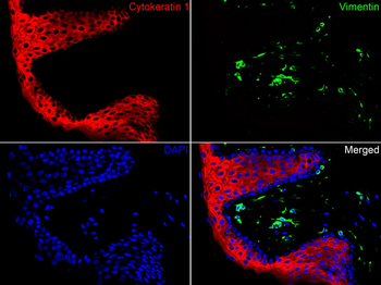
Immunofluorescence analysis of paraffin-embedded human skin tissue labeling Cytokeratin 1 (orb559083) and Vimentin. The section was pre-treated using heat mediated antigen retrieval with Tris-EDTA buffer (pH9.0) for 20 minutes. The tissues were blocked in 10% negative goat serum for 1 hour at room temperature, washed with PBS. And then probed with the primary antibodies Cytokeratin 1 (orb559083, red) at 1/200 dilution and Vimentin (green) at 1/200 dilution at +4C overnight, washed with PBS. |
|
|
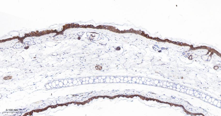
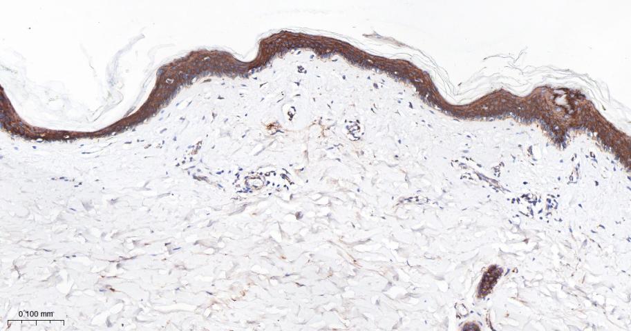
Immunohistochemical analysis of paraffin-embedded human skin tissue using anti-Cytokeratin 1 antibody. The section was pre-treated using heat mediated antigen retrieval with Tris-EDTA buffer (pH9.0) for 20 minutes. The tissues were blocked in 5% BSA for 30 minutes at room temperature, washed with ddH2O and PBS, and then probed with the primary antibody (orb559083, 1/400) for 30 minutes at room temperature. The detection was performed using an HRP conjugated compact polymer system. DAB was used as the chromogen. Tissues were counterstained with hematoxylin and mounted with DPX. |
|
|
Immunohistochemical analysis of paraffin-embedded mouse skin tissue using anti-Cytokeratin 1 antibody. The section was pre-treated using heat mediated antigen retrieval with Tris-EDTA buffer (pH 8.0-8.4) for 20 minutes. The tissues were blocked in 5% BSA for 30 minutes at room temperature, washed with ddH2O and PBS, and then probed with the primary antibody (orb559083, 1/50) for 30 minutes at room temperature. The detection was performed using an HRP conjugated compact polymer system. DAB was used as the chromogen. Tissues were counterstained with hematoxylin and mounted with DPX. |
|
|
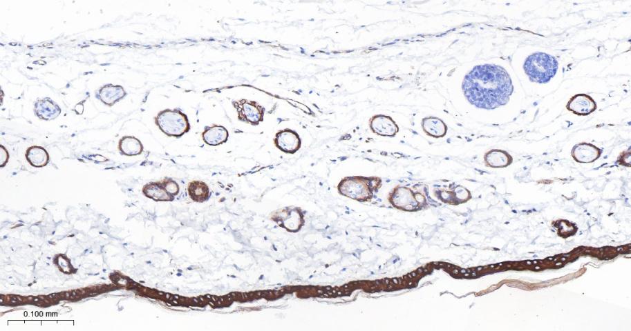
Immunohistochemical analysis of paraffin-embedded rat skin tissue with Rabbit anti-Cytokeratin 1 antibody (orb559083) at 1/400 dilution. The section was pre-treated using heat mediated antigen retrieval with Tris-EDTA buffer (pH9.0) for 20 minutes. The tissues were blocked in 1% BSA for 20 minutes at room temperature, washed with ddH2O and PBS, and then probed with the primary antibody (orb559083) at 1/400 dilution for 1 hour at room temperature. The detection was performed using an HRP conjugated compact polymer system. DAB was used as the chromogen. Tissues were counterstained with hematoxylin and mounted with DPX. |
|
|
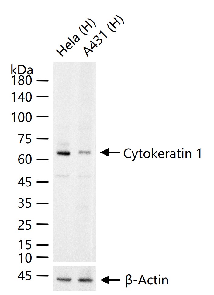
Western blot analysis of Cytokeratin 1 on different lysates. Proteins were transferred to a PVDF membrane and blocked with 5% BSA in PBS for 1 hour at room temperature. The primary antibody (orb559083, 1/500) was used in 5% BSA at room temperature for 2 hours. Goat Anti-Rabbit IgG - HRP Secondary Antibody at 1:5000 dilution was used for 1 hour at room temperature. Positive control: Lane 1: Hela cell lysate, Lane 2: 293T cell lysate. |
|
|
25 ug total protein per lane of various lysates probed with Cytokeratin 1 Recombinant Rabbit Monoclonal Antibody (orb559083) at 1:1000 dilution and 4C overnight incubation. Followed by conjugated secondary antibody incubation at r.t. for 60 min. |
|
|
Paraformaldehyde-fixed, paraffin embedded Mouse Skin, Antigen retrieval by boiling in sodium citrate buffer (pH6.0) for 15 min, Antibody incubation with Cytokeratin 1 Recombinant Rabbit Monoclonal Antibody (orb559083) at 1:200 overnight at 4C, followed by conjugation to the Goat Anti-Rabbit IgG H&L, HRP conjugated (orb572747) and DAB staining. |
|
|
Paraformaldehyde-fixed, paraffin embedded Rat Skin, Antigen retrieval by boiling in sodium citrate buffer (pH6.0) for 15 min, Cytokeratin 1 Recombinant Rabbit Monoclonal Antibody (orb559083) at 1:200 overnight at 4C, followed by conjugation to the Goat Anti-Rabbit IgG H&L, HRP conjugated (orb572747) and DAB staining. |
|
|
Paraformaldehyde-fixed, paraffin embedded Human Skin, Antigen retrieval by boiling in sodium citrate buffer (pH6.0) for 15 min, Cytokeratin 1 Recombinant Rabbit Monoclonal Antibody (orb559083) at 1:200 overnight at 4C, followed by conjugation to the Goat Anti-Rabbit IgG H&L, HRP conjugated (orb572747) and DAB staining. |
Produktgarantie und fachkundiger Support
