
IRF1 Recombinant Rabbit Monoclonal Antibody, Clone: [3G7], Unconjugated
Artikelnummer:
BYT-ORB559092
- Bilder (9)


| Artikelname: | IRF1 Recombinant Rabbit Monoclonal Antibody, Clone: [3G7], Unconjugated |
| Artikelnummer: | BYT-ORB559092 |
| Hersteller Artikelnummer: | orb559092 |
| Alternativnummer: | BYT-ORB559092-50,BYT-ORB559092-100 |
| Hersteller: | Biorbyt |
| Wirt: | Rabbit |
| Kategorie: | Antikörper |
| Applikation: | FC, ICC, IF, IHC-Fr, IHC-P, WB |
| Spezies Reaktivität: | Human |
| Immunogen: | Recombinant human IRF1 protein, around C-terminal 100aa |
| Konjugation: | Unconjugated |
| Alternative Synonym: | IMD117, IRF-1, MAR, IRF1_HUMAN, IRF1, |
| IRF1 Recombinant Rabbit Monoclonal Antibody |
| Klonalität: | Recombinant |
| Konzentration: | 1mg/ml |
| Klon-Bezeichnung: | [3G7] |
| Molekulargewicht: | 37 kDa |
| UniProt: | P10914 |
| Puffer: | 0.01M TBS (pH7.4) with 1% rAlbumin, 0.02% Proclin300 and 50% Glycerol. |
| Formulierung: | Liquid |
| Target-Kategorie: | IRF1 |
| Application Verdünnung: | WB=1:500-2000, IHC-P=1:50-200, IHC-F=1:50-200, ICC/IF=1:50-200, IF=1:50-200, Flow-Cyt=1:50-100 |
|
|
Blank control: Jurkat. Primary Antibody (green line): Rabbit Anti-IRF1 antibody (orb559092), dilution: 1:50, Secondary Antibody: Goat anti-rabbit IgG-AF488, dilution: 1:1000. Protocol, The cells were fixed with 4% PFA (10 min at room temperature) and then permeabilized with 90% ice-cold methanol for 20 min at -20C. The cells were then incubated in 5% BSA to block non-specific protein-protein interactions for 30 min at room temperature. Cells stained with Primary Antibody for 30 min at room temperature. The secondary antibody used for 40 min at room temperature. Acquisition of 20000 events was performed. |
|
|
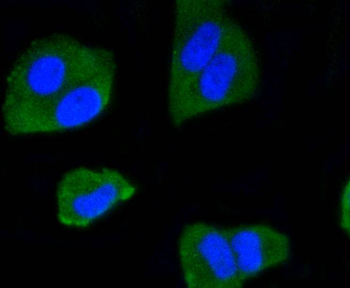
Hela cell, 4% Paraformaldehyde-fixed, Triton X-100 at room temperature for 20 min, Blocking buffer (normal goat serum) at 37C for 20 min, Antibody incubation with (IRF1) monoclonal Antibody, Unconjugated (orb559092) 1:50, 90 minutes at 37C, followed by a conjugated Goat Anti-Rabbit IgG antibody at 37C for 90 minutes, DAPI (blue) was used to stain the cell nuclei. |
|
|
NIH/3T3 cell, 4% Paraformaldehyde-fixed, Triton X-100 at room temperature for 20 min, Blocking buffer (normal goat serum) at 37C for 20 min, Antibody incubation with (IRF1) monoclonal Antibody, Unconjugated (orb559092) 1:50, 90 minutes at 37C, followed by a conjugated Goat Anti-Rabbit IgG antibody at 37C for 90 minutes, DAPI (blue) was used to stain the cell nuclei. |
|
|
Paraformaldehyde-fixed, paraffin embedded (human breast carcinoma), Antigen retrieval by boiling in sodium citrate buffer (pH6.0) for 15 min, Block endogenous peroxidase by 3% hydrogen peroxide for 20 minutes, Blocking buffer (normal goat serum) at 37C for 30 min, Antibody incubation with (IRF1) Monoclonal Antibody, Unconjugated (orb559092) at 1:50 overnight at 4C, followed by operating according to SP Kit (Rabbit) instructionsand DAB staining. |
|
|
Paraformaldehyde-fixed, paraffin embedded (human colon carcinoma), Antigen retrieval by boiling in sodium citrate buffer (pH6.0) for 15 min, Block endogenous peroxidase by 3% hydrogen peroxide for 20 minutes, Blocking buffer (normal goat serum) at 37C for 30 min, Antibody incubation with (IRF1) Monoclonal Antibody, Unconjugated (orb559092) at 1:50 overnight at 4C, followed by operating according to SP Kit (Rabbit) instructionsand DAB staining. |
|
|
Paraformaldehyde-fixed, paraffin embedded (mouse brain), Antigen retrieval by boiling in sodium citrate buffer (pH6.0) for 15 min, Block endogenous peroxidase by 3% hydrogen peroxide for 20 minutes, Blocking buffer (normal goat serum) at 37C for 30 min, Antibody incubation with (IRF1) Monoclonal Antibody, Unconjugated (orb559092) at 1:50 overnight at 4C, followed by operating according to SP Kit (Rabbit) instructionsand DAB staining. |
|
|
Sample: Lane 1: Human THP-1 cell lysates, Lane 2: Human Jurkat cell lysates, Lane 3: Human MOLT4 cell lysates, Primary: Anti-IRF1 (orb559092) at 1/1000 dilution, Secondary: IRDye800CW Goat Anti-Rabbit IgG at 1/20000 dilution, Predicted band size: 37 kDa, Observed band size: 46 kDa. |
|
|
Sample: Lane 1: PC-12 cell lysates, Lane 2: Jurkat cell lysates, Primary: Anti-IRF1 (orb559092) at 1/500 dilution, Secondary: Goat Anti-Rabbit IgG - HRP at 1/5000 dilution, Predicted band size: 37 kD, Observed band size: 48 kD. |
|
|
Western blot:Lane 1:PC-12 cell,Lane 2:Jurkat cell,Primary:Anti-IRF1 at 1:500 dilution. |
Produktgarantie und fachkundiger Support
