
Histone H4 Recombinant Rabbit Monoclonal Antibody, Clone: [2G2], Unconjugated
Artikelnummer:
BYT-ORB559095
- Bilder (9)


| Artikelname: | Histone H4 Recombinant Rabbit Monoclonal Antibody, Clone: [2G2], Unconjugated |
| Artikelnummer: | BYT-ORB559095 |
| Hersteller Artikelnummer: | orb559095 |
| Alternativnummer: | BYT-ORB559095-50,BYT-ORB559095-100 |
| Hersteller: | Biorbyt |
| Wirt: | Rabbit |
| Kategorie: | Antikörper |
| Applikation: | FC, IF, IHC-Fr, IHC-P, WB |
| Spezies Reaktivität: | Human, Mouse, Rat |
| Immunogen: | A synthesized peptide derived from human Histone H4 (80-103/103aa) |
| Konjugation: | Unconjugated |
| Alternative Synonym: | H4-16, H4/p, H4C1, H4C11, H4C12, H4C13, H4C14, H4C15, H4C2, H4C3, H4C4, H4C5, H4C6, H4C8, H4C9, HIST4H4, H4/o, H4C16, HIST2H4B, H4/m, H4FM, H4M, HIST1H4I, TEBIVANED4, TEVANED4, H4FA, HIST1H4A, H4/b, H4FB, HIST1H4D, dJ221C16.9, H4, H4/c, H4FC, HIST1H4F, H4/d, H4F2iii, H4FD, HIST1H4K, dJ160A22.1, H4/e, H4F2iv, H4FE, HIST1H4J, TEBIVANED2, TEVANED2, dJ160A22.2, H4/g, H4FG, HIST1H4C, TEBIVANED1, TEVANED1, dJ221C16.1, H4/h, H4FH, HIST1H4H, H4/I, H4FI, HIST1H4B, H4/j, H4FJ, HIST1H4E, TEBIVANED3, TEVANED3, H4.k, H4/k, H4FK, HIST1H4L, FO108, H4/n, H4F2, H4FN, HIST2H4, HIST2H4A, Gm11275, H4f16, Hist1h4m, Hist1h4n, B130044J01Rik, 1700024H08Rik, H2c8, H4_DROME, His4, His4r, His4:CG31611, His4:CG33869, His4:CG33871, His4:CG33873, His4:CG33875, His4:CG33877, His4:CG33879, His4:CG33881, His4:CG33883, His4:CG33885, His4:CG33887, His4:CG33889, His4:CG33891, His4:CG33893, His4:CG33895, His4:CG33897, His4:CG33899, His4:CG33901, His4:CG33903, His4:CG33905, His4:CG33907, His4:CG33909, H4r, H4_HUMAN, H4/A, H4FO, H4_MOUSE, H4-53, H4-12, H4_SCHPO, hhf1, hhf2, hhf3, h4.1, h4.2, h4.3, |
| Histone H4 Recombinant Rabbit Monoclonal Antibody |
| Klonalität: | Recombinant |
| Konzentration: | 1mg/ml |
| Klon-Bezeichnung: | [2G2] |
| Molekulargewicht: | 11 kDa |
| UniProt: | P62805 |
| Puffer: | 0.01M TBS (pH7.4) with 1% rAlbumin, 0.02% Proclin300 and 50% Glycerol. |
| Formulierung: | Liquid |
| Target-Kategorie: | H4C1 |
| Application Verdünnung: | WB=1:500-2000, IHC-P=1:200-1000, IHC-F=1:200-1000, IF=1:200-1000, Flow-Cyt=1ug/Test |
|
|
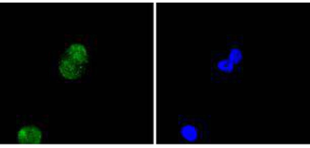
ICC staining of Histone H4 in PANC-1 cells (green). Formalin fixed cells were permeabilized with 0.1% Triton X-100 in TBS for 10 minutes at room temperature and blocked with 1% Blocker BSA for 15 minutes at room temperature. Cells were probed with the primary antibody (orb559095, 1/50) for 1 hour at room temperature, washed with PBS. Alexa Fluor488 Goat anti-Rabbit IgG was used as the secondary antibody at 1/1000 dilution. The nuclear counter stain is DAPI (blue). |
|
|
Immunohistochemical analysis of paraffin-embedded human breast carcinoma tissue using anti-Histone H4 antibody. The section was pre-treated using heat mediated antigen retrieval with Tris-EDTA buffer (pH 8.0-8.4) for 20 minutes. The tissues were blocked in 5% BSA for 30 minutes at room temperature, washed with ddH2O and PBS, and then probed with the primary antibody (1/50) for 30 minutes at room temperature. The detection was performed using an HRP conjugated compact polymer system. DAB was used as the chromogen. Tissues were counterstained with hematoxylin and mounted with DPX. |
|
|
Immunohistochemical analysis of paraffin-embedded human kidney tissue with Rabbit anti-Histone H4 antibody (orb559095) at 1/5000 dilution. The section was pre-treated using heat mediated antigen retrieval with sodium citrate buffer (pH 6.0) for 2 minutes. The tissues were blocked in 1% BSA for 20 minutes at room temperature, washed with ddH2O and PBS, and then probed with the primary antibody (orb559095) at 1/5000 dilution for 1 hour at room temperature. The detection was performed using an HRP conjugated compact polymer system. DAB was used as the chromogen. Tissues were counterstained with hematoxylin and mounted with DPX. |
|
|
Immunohistochemical analysis of paraffin-embedded human pancreas tissue with Rabbit anti-Histone H4 antibody (orb559095) at 1/5000 dilution. The section was pre-treated using heat mediated antigen retrieval with sodium citrate buffer (pH 6.0) for 2 minutes. The tissues were blocked in 1% BSA for 20 minutes at room temperature, washed with ddH2O and PBS, and then probed with the primary antibody (orb559095) at 1/5000 dilution for 1 hour at room temperature. The detection was performed using an HRP conjugated compact polymer system. DAB was used as the chromogen. Tissues were counterstained with hematoxylin and mounted with DPX. |
|
|
Immunohistochemical analysis of paraffin-embedded mouse colon tissue using anti-Histone H4 antibody. The section was pre-treated using heat mediated antigen retrieval with Tris-EDTA buffer (pH 8.0-8.4) for 20 minutes. The tissues were blocked in 5% BSA for 30 minutes at room temperature, washed with ddH2O and PBS, and then probed with the primary antibody (orb559095, 1/50) for 30 minutes at room temperature. The detection was performed using an HRP conjugated compact polymer system. DAB was used as the chromogen. Tissues were counterstained with hematoxylin and mounted with DPX. |
|
|
Immunohistochemical analysis of paraffin-embedded mouse skin tissue using anti-Histone H4 antibody. The section was pre-treated using heat mediated antigen retrieval with Tris-EDTA buffer (pH 8.0-8.4) for 20 minutes. The tissues were blocked in 5% BSA for 30 minutes at room temperature, washed with ddH2O and PBS, and then probed with the primary antibody (orb559095, 1/50) for 30 minutes at room temperature. The detection was performed using an HRP conjugated compact polymer system. DAB was used as the chromogen. Tissues were counterstained with hematoxylin and mounted with DPX. |
|
|
Immunohistochemical analysis of paraffin-embedded mouse testis tissue with Rabbit anti-Histone H4 antibody (orb559095) at 1/5000 dilution. The section was pre-treated using heat mediated antigen retrieval with sodium citrate buffer (pH 6.0) for 2 minutes. The tissues were blocked in 1% BSA for 20 minutes at room temperature, washed with ddH2O and PBS, and then probed with the primary antibody (orb559095) at 1/5000 dilution for 1 hour at room temperature. The detection was performed using an HRP conj |
|
|
|
|
|
Produktgarantie und fachkundiger Support
