136.36mM Ethanolamine, 133.23 mM Chlorides, 9.55mM Phosphates, 9.55mM Sodium Bicarbonate
Target-Kategorie:
VPS35
Application Verdünnung:
WB (1:1000), ICC/IF (1:200), IP (1:200)
Anwendungsbeschreibung:
Application Notes: A 1:1000 dilution of SMC-602 was sufficient for detection of VPS35 in 10 µg of SH-SY5Y by ECL immunoblot analysis using Goat Anti-Mouse IgG:HRP as the secondary antibody
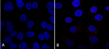
Immunocytochemistry/Immunofluorescence analysis using Mouse Anti-VPS35 Monoclonal Antibody, Clone 7E4. Tissue: A549 cells. Species: Human. Primary Antibody: Mouse Anti-VPS35 Monoclonal Antibody at 1:5 (tissue culture supernatant). Secondary Antibody: Donkey anti-mouse: Alexa Fluor 594 at 1:4000 in 0.2% BSA PBS. Counterstain: DAPI. Localization: Vesicles. A) VPS35 KO A549 cells B) WT A549 cells.
Immunoprecipitation analysis using Mouse Anti-VPS35 Monoclonal Antibody, Clone 7E4. Tissue: A549 cells. Species: Human. Primary Antibody: Mouse Anti-VPS35 Monoclonal Antibody. 500 µL cell culture supernatants were incubated with 10 µL of Protein A/G resin beads for 1 hour at 4C. clone 7E4 depletes virtually all of the VPS35 from the A549 cell extract.
Western Blot analysis of Human SH-SY5Y lysates showing detection of 91.7 kDa VPS35 protein using Mouse Anti-VPS35 Monoclonal Antibody, Clone 7E4. Lane 1: Molecular Weight Ladder. Lane 2: SH-SY5Y. Load: 10 µg. Block: 5% Skim Milk powder in TBST. Primary Antibody: Mouse Anti-VPS35 Monoclonal Antibody at 1:1000 for 2 hours at RT with shaking. Secondary Antibody: Goat anti-mouse IgG:HRP at 1:4000 for 1 hour at RT with shaking. Color Development: Chemiluminescent for HRP (Moss) for 5 min in RT. Predicted/Observed Size: 91.7 kDa.
Immunoprecipitation analysis using Mouse Anti-VPS35 Monoclonal Antibody, Clone 7E4. Tissue: A549 cells. Species: Human. Primary Antibody: Mouse Anti-VPS35 Monoclonal Antibody. Three amounts of (3, 1 and 0.3 ug) were non-covalently coupled to 10uL of A/G sepharose beads for 1 hour at 4C and next incubated with 250ug of A549 lysate for 2 hours at 4C.
Immunoprecipitation analysis using Mouse Anti-VPS35 Monoclonal Antibody, Clone 7E4. Tissue: embryonic fibroblast. Species: Mouse. Primary Antibody: Mouse Anti-VPS35 Monoclonal Antibody. Three amounts of (3, 1 and 0.3 ug) were non-covalently coupled to 10uL of A/G sepharose beads for 1 hour at 4C and next incubated with 250ug of MEF lysate for 2 hours at 4C.
Immunoprecipitation analysis using Mouse Anti-VPS35 Monoclonal Antibody, Clone 7E4. Tissue: MEF, A549 cells. Species: Human, Mouse. Primary Antibody: Mouse Anti-VPS35 Monoclonal Antibody at 1:5 (tissue culture supernatant). 10 ug antibody were coupled to 10 uL A/G resin beads either covalently (with DMP) or non-covalently (1 hour at 4 degrees). The antibody immunoprecipitates VPS35 in mouse and human cells effectively when covalently coupled to the beads.
Western Blot analysis of Human, Mouse A549, MEF showing detection of VPS35 protein using Mouse Anti-VPS35 Monoclonal Antibody, Clone 7E4. Lane 1: Molecular Weight Ladder. Lane 2: VPS35 KO A549 cells. Lane 3: mouse embryonic fibroblast cells. Load: 8 µg each A549 and MEF. Primary Antibody: Mouse Anti-VPS35 Monoclonal Antibody at 1:5 (tissue culture supernatant). Secondary Antibody: Donkey anti-mouse IRDye 800CW at 1:25000 in TBS-T.
* Mehrwertsteuer und Versandkosten nicht enthalten. Irrtümer und Preisänderungen vorbehalten