
Phospho-TAK1 (Thr187) Rabbit Polyclonal Antibody, Unconjugated
Artikelnummer:
BYT-ORB7049
- Bilder (9)


| Artikelname: | Phospho-TAK1 (Thr187) Rabbit Polyclonal Antibody, Unconjugated |
| Artikelnummer: | BYT-ORB7049 |
| Hersteller Artikelnummer: | orb7049 |
| Alternativnummer: | BYT-ORB7049-50,BYT-ORB7049-100,BYT-ORB7049-200 |
| Hersteller: | Biorbyt |
| Wirt: | Rabbit |
| Kategorie: | Antikörper |
| Applikation: | FC, IF, IHC-Fr, IHC-P, WB |
| Spezies Reaktivität: | Human, Mouse, Rat |
| Immunogen: | KLH conjugated Synthesised phosphopeptide derived from human TAK1 around the phosphorylation site of Thr187 HM(p-T)NN |
| Konjugation: | Unconjugated |
| Alternative Synonym: | MAP3K7 | TAK1 (p-T187), p-TAK1, phospho-TAK1, CSCF, FMD2, MEKK7, TAK1, TGF1a, B430101B05, M3K7_HUMAN, MAP3K7, Transforming growth factor-beta-activated kinase 1 (TGF-beta-activated kinase 1), 2.7.11.25, M3K7_MOUSE, M3K7_RAT, |
| Phospho-TAK1 (Thr187) Rabbit Polyclonal Antibody |
| Application Verdünnung: | WB=1:500-2000, IHC-P=1:100-500, IHC-F=1:100-500, IF=1:100-500, Flow-Cyt=1µg/Test |
| Anwendungsbeschreibung: | Modification: Phosphorylated |
|
|
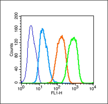
Blank control (Black line): Raji (Black). Primary Antibody (green line): Rabbit Anti-Phospho-TAK1 (Thr187) antibody (orb7049), dilution: 1 µg/10 6 cells, Isotype Control Antibody (orange line): Rabbit IgG. Secondary Antibody (white blue line): Goat anti-rabbit IgG-PE, dilution: 1 µg/Test. Protocol, The cells were fixed with 70% ice-cold methanol overnight at 4C and then permeabilized with 0.1% PBS-Tween for 20 min at room temperature (The cells were fixed with 2% paraformaldehyde (10 min), then permeabilized with 90% ice-cold methanol for 20 min on ice.). Cells stained with Primary Antibody for 30 min at room temperature. The cells were then incubated in 1X PBS/2% BSA/10% goat serum to block non-specific protein-protein interactions followed by the antibody for 15 min at room temperature. The secondary antibody used for 40 min at room temperature. Acquisition of 20000 events was performed. |
|
|
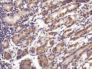
Paraformaldehyde-fixed, paraffin embedded (human gastric carcinoma), Antigen retrieval by boiling in sodium citrate buffer (pH6.0) for 15 min, Block endogenous peroxidase by 3% hydrogen peroxide for 20 minutes, Blocking buffer (normal goat serum) at 37C for 30 min, Antibody incubation with (Phospho-TAK1 (Thr187)) Polyclonal Antibody, Unconjugated (orb7049) at 1:200 overnight at 4C, followed by operating according to SP Kit (Rabbit) instructionsand DAB staining. |
|
|
Paraformaldehyde-fixed, paraffin embedded (Human stomach), Antigen retrieval by microwave in sodium citrate buffer (pH6.0), Block endogenous peroxidase by 3% hydrogen peroxide for 30 minutes, Blocking buffer (3% BSA) at RT for 30 min, Antibody incubation with (Phospho-TAK1 (Thr187)) Polyclonal Antibody, Unconjugated (orb7049) at 1:400 overnight at 4C, followed by conjugation to the secondary antibody (labeled with HRP) and DAB staining. |
|
|
Paraformaldehyde-fixed, paraffin embedded (Mouse brain), Antigen retrieval by microwave in sodium citrate buffer (pH6.0), Block endogenous peroxidase by 3% hydrogen peroxide for 30 minutes, Blocking buffer (3% BSA) at RT for 30 min, Antibody incubation with (Phospho-TAK1 (Thr187)) Polyclonal Antibody, Unconjugated (orb7049) at 1:400 overnight at 4C, followed by conjugation to the secondary antibody (labeled with HRP) and DAB staining. |
|
|
Paraformaldehyde-fixed, paraffin embedded (Rat brain), Antigen retrieval by microwave in sodium citrate buffer (pH6.0), Block endogenous peroxidase by 3% hydrogen peroxide for 30 minutes, Blocking buffer (3% BSA) at RT for 30 min, Antibody incubation with (Phospho-TAK1 (Thr187)) Polyclonal Antibody, Unconjugated (orb7049) at 1:400 overnight at 4C, followed by conjugation to the secondary antibody (labeled with HRP) and DAB staining. |
|
|
Sample: Lane 1: MCF-7 (Human) Cell Lysate at 30 ug, Lane 2: Hela (Human) Cell Lysate at 30 ug, Lane 3: A431 (Human) Cell Lysate at 30 ug, Lane 4: 293T (Human) Cell Lysate at 30 ug, Lane 5: K562 (Human) Cell Lysate at 30 ug, Primary: Anti-Phospho-TAK1 (Thr187) (orb7049) at 1/1000 dilution, Secondary: IRDye800CW Goat Anti-Rabbit IgG at 1/20000 dilution, Predicted band size: 78 kD, Observed band size: 75 kD. |
|
|
Sample: Muscle (Mouse) Lysate at 40 ug, Primary: Anti-Phospho-TAK1 (Thr187) (orb7049) at 1/1000 dilution, Secondary: IRDye800CW Goat Anti-Rabbit IgG at 1/20000 dilution, Predicted band size: 67 kD, Observed band size: 70 kD. |
|
|
IHC-P analysis of Human stomach using Phospho-TAK1 Thr187 Polyclonal Antibody at 1:400 dilution. |
|
|
IHC-P analysis of Mouse brain using Phospho-TAK1 Thr187 Polyclonal Antibody at 1:400 dilution. |
Produktgarantie und fachkundiger Support
