Western blot, 0.1-0.25 µg/ml, Human, Mouse Immunohistochemistry(Paraffin-embedded Section), 1-2 µg/ml, Human
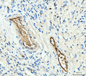
IHC analysis of Hemoglobin/HBA1/HBA2 using anti-Hemoglobin/HBA1/HBA2 antibody. Hemoglobin/HBA1/HBA2 was detected in a paraffin-embedded section of human gastric signet ring cell carcinoma tissue. Heat mediated antigen retrieval was performed in EDTA buffer (pH8.0, epitope retrieval solution). The tissue section was blocked with 10% goat serum. The tissue section was then incubated with 2 µg/ml rabbit anti-Hemoglobin/HBA1/HBA2 Antibody overnight at 4C. Biotinylated goat anti-rabbit IgG was used as secondary antibody and incubated for 30 minutes at 37C. The tissue section was developed using Strepavidin-Biotin-Complex (SABC) with DAB as the chromogen.
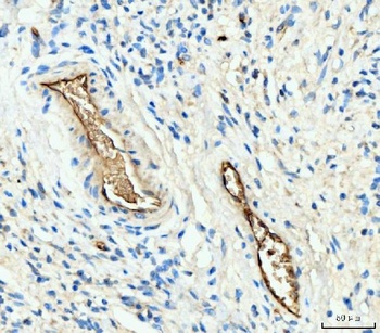
IHC analysis of Hemoglobin/HBA1/HBA2 using anti-Hemoglobin/HBA1/HBA2 antibody. Hemoglobin/HBA1/HBA2 was detected in a paraffin-embedded section of human rectal moderately differentiated adenocarcinoma tissue. Heat mediated antigen retrieval was performed in EDTA buffer (pH8.0, epitope retrieval solution). The tissue section was blocked with 10% goat serum. The tissue section was then incubated with 2 µg/ml rabbit anti-Hemoglobin/HBA1/HBA2 Antibody overnight at 4C. Biotinylated goat anti-rabbit IgG was used as secondary antibody and incubated for 30 minutes at 37C. The tissue section was developed using Strepavidin-Biotin-Complex (SABC) with DAB as the chromogen.
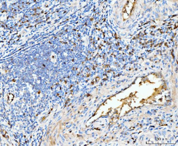
IHC analysis of Hemoglobin/HBA1/HBA2 using anti-Hemoglobin/HBA1/HBA2 antibody. Hemoglobin/HBA1/HBA2 was detected in a paraffin-embedded section of human renal cancer tissue. Heat mediated antigen retrieval was performed in EDTA buffer (pH8.0, epitope retrieval solution). The tissue section was blocked with 10% goat serum. The tissue section was then incubated with 2 µg/ml rabbit anti-Hemoglobin/HBA1/HBA2 Antibody overnight at 4C. Biotinylated goat anti-rabbit IgG was used as secondary antibody and incubated for 30 minutes at 37C. The tissue section was developed using Strepavidin-Biotin-Complex (SABC) with DAB as the chromogen.
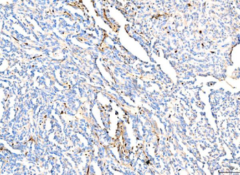
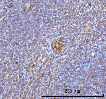
IHC analysis of Hemoglobin/HBA1/HBA2 using anti-Hemoglobin/HBA1/HBA2 antibody. Hemoglobin/HBA1/HBA2 was detected in a paraffin-embedded section of human spleen tissue. Heat mediated antigen retrieval was performed in EDTA buffer (pH8.0, epitope retrieval solution). The tissue section was blocked with 10% goat serum. The tissue section was then incubated with 2 µg/ml rabbit anti-Hemoglobin/HBA1/HBA2 Antibody overnight at 4C. Biotinylated goat anti-rabbit IgG was used as secondary antibody and incubated for 30 minutes at 37C. The tissue section was developed using Strepavidin-Biotin-Complex (SABC) with DAB as the chromogen.
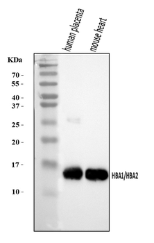
Western blot analysis of Hemoglobin/HBA1/HBA2 using anti-Hemoglobin/HBA1/HBA2 antibody. Electrophoresis was performed on a 5-20% SDS-PAGE gel at 70V (Stacking gel) / 90V (Resolving gel) for 2-3 hours. The sample well of each lane was loaded with 30 ug of sample under reducing conditions. Lane 1: human placenta tissue lysates, Lane 2: mouse heart tissue lysates. After electrophoresis, proteins were transferred to a nitrocellulose membrane at 150 mA for 50-90 minutes. Blocked the membrane with 5% non-fat milk/TBS for 1.5 hour at RT. The membrane was incubated with rabbit anti-Hemoglobin/HBA1/HBA2 antigen affinity purified polyclonal antibody at 0.25 µg/mL overnight at 4C, then washed with TBS-0.1% Tween 3 times with 5 minutes each and probed with a goat anti-rabbit IgG-HRP secondary antibody at a dilution of 1:5000 for 1.5 hour at RT. The signal is developed using an Enhanced Chemiluminescent detection (ECL) kit with Tanon 5200 system. A specific band was detected for Hemoglobin/HBA1/HBA2 at approximately 15 kDa. The expected band size for Hemoglobin/HBA1/HBA2 is at 15 kDa.
* Mehrwertsteuer und Versandkosten nicht enthalten. Irrtümer und Preisänderungen vorbehalten