
FLAER (Alexa 488 proaerolysin variant) liquid format-IVD
- Bilder (1)
| Artikelname: | FLAER (Alexa 488 proaerolysin variant) liquid format-IVD |
| Artikelnummer: | CED-FL1S-C |
| Hersteller Artikelnummer: | FL1S-C |
| Alternativnummer: | CED-FL1S-C |
| Hersteller: | Cedarlane |
| Kategorie: | Sonstiges |
| FLAER (Alexa 488 proaerolysin variant) liquid format-IVD |
For In Vitro Diagnostic Use.
Intended UseFLAER is used in a clinical laboratory setting for multi-parameter flow cytometry along with antibodies (including CD45, CD33, CD24, CD15 and CD14) to detect PNH clones (FLAER-negative cells) within the monocyte and granulocyte (cell) lineages. 6,7,9 The result is a sensitive an accurate test that can be used in combination with the CD55/CD59 assay for detecting PHN clones in red blood cells. Paroxysmal nocturnal hemoglobinuria (PNH) is a stem cell disorder caused by a mutation of the gene involved in the synthesis of the GPI (glycosylphosphatidylinositol) anchor of a group of surface proteins on circulating cells. Affected cells are sensitive to complement-mediated hemolysis and this may lead to life-threatening thrombosis, chronic kidney disease, pulmonary hypertension, end organ damage, ischemic bowel disease, hepatic failure, and anemia. Identifying PNH patients early in the course of their disease may offer the best opportunity for long-term management. In the past, PNH has been challenging to identify effectively. However, in recent years impressive strides have been made in the understanding of PNH pathology, accompanied by greatly improved detection techniques, including multiparametric flow cytometry using FLAER.
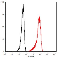
About FLAERFLAER is an Alexa Fluor® 488 labeled variant of aerolysin, a unique protein that binds tightly and specifically to mammalian GPI anchors. FLAER will not bind to PNH cells because they do not produce the anchor. Before FLAER, detection of PNH clones by flow cytometry relied on fluorescently labeled antibodies to GPI-linked proteins such as CD59 and CD55. These antibodies do not bind with high affinity, so that small PNH clones are not detected. Also, they each screen for the absence of a specific protein, rather than loss of the GPI anchor, and therefore there is the risk of false negative results. Since FLAER binds to the GPI anchor itself, only PNH cells, which lack the anchor, will be negative. And since FLAER binds with high affinity, very small PNH populations can be detected. Formulation: FL1S-C contains 25 μg of FLAER in 0.5 ml PBS-albumin (50 tests), 0.05% Sodium Azide. FL2S-C contains 50 μg of FLAER in 1.0 ml PBS-albumin (100 tests), 0.05% Sodium Azide The concentration of FLAER in the supplied stock solution is 10 -6 M. The solution also contains 5 mg/mL albumin, added in order to maximize stability. Performance: On healthy individuals, FLAER binds to essentially all human lymphocytes, monocytes and granulocytes. In our testing, lymphocytes were labelled with FLAER as described above. The histogram below outlines count versus fluorescence intensity of FLAER labeled lymphocytes from a healthy individual (acquisition is performed using the Accuri C6 and analyzed in FCS Express). FLAER (red histogram) at a dilution of 5.0x10⁻⁸ M vs. unstained cells (black histogram).
|

| Lagerung: | 2°C to 8°C |
|
|
FLAER (red histogram) at a dilution of 5.0x10⁻⁸ M vs. unstained cells (black histogram). |
