
HSPA8 Monoclonal Antibody, IgG2b, Clone: [2G8F6], Unconjugated, Mouse
Artikelnummer:
CSB-MA010829A0M
- Bilder (7)
| Artikelname: | HSPA8 Monoclonal Antibody, IgG2b, Clone: [2G8F6], Unconjugated, Mouse |
| Artikelnummer: | CSB-MA010829A0M |
| Hersteller Artikelnummer: | CSB-MA010829A0m |
| Alternativnummer: | CSB-MA010829A0M-100UL, CSB-MA010829A0M-50UL |
| Hersteller: | Cusabio |
| Wirt: | Mouse |
| Kategorie: | Antikörper |
| Applikation: | ELISA, FC, IHC, IP, WB |
| Spezies Reaktivität: | Human, Mouse, Rat |
| Konjugation: | Unconjugated |
| Alternative Synonym: | Heat shock cognate 71 kDa protein (Heat shock 70 kDa protein 8) (Lipopolysaccharide-associated protein 1) (LAP-1) (LPS-associated protein 1), HSPA8, HSC70 HSP73 HSPA10 |
| Klonalität: | Monoclonal |
| Klon-Bezeichnung: | [2G8F6] |
| Isotyp: | IgG2b |
| UniProt: | P11142 |
| Puffer: | Preservative: 0.03% Proclin 300<br />Constituents: 50% Glycerol, 0.01M PBS, PH 7.4 |
| Reinheit: | >95%, Protein A purified |
| Formulierung: | Liquid |
| Target-Kategorie: | HSPA8 |
| Application Verdünnung: | Recommended dilution: WB: 1:10000-1:256000, IHC: 1:100-1:500, FC: 1:100-1:300, IP: 1µl-4µl |
|
|
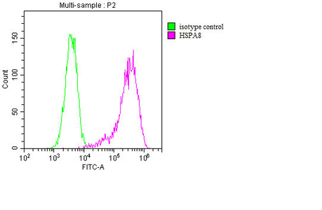
Overlay histogram showing MCF-7 cells stained with CSB-MA010829A0m (red line). The cells were fixed with 70% Ethylalcohol (18h) and then incubated in 10% normal goat serum to block non-specific protein-protein interactions followed by the primary antibody at 1/200 for 1 h at 4C. The secondary antibody used was FITC goat anti-mouse IgG(H+L) at 1/100 dilution for 30min at 4C. Isotype control antibody (green line) was mouse IgG2b used under the same conditions. Acquisition of >10,000 events was performed. |
|
|
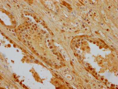
IHC image of CSB-MA010829A0m diluted at 1:256 and staining in paraffin-embedded human prostate cancer performed on a Leica BondTM system. After dewaxing and hydration, antigen retrieval was mediated by high pressure in a citrate buffer (pH 6.0). Section was blocked with 10% normal goat serum 30min at RT. Then primary antibody (1% BSA) was incubated at 4C overnight, and detected by a Goat anti-mouse IgG polymer labeled by HRP and visualized using 0.05% DAB. |
|
|
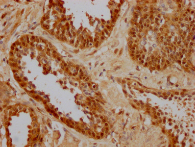
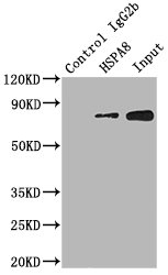
Immunoprecipitating HSPA8 in Hela whole cell lysate Lane 1: Mouse control IgG2b instead of CSB-MA010829A0m in Hela whole cell lysate.Lane 2: CSB-MA010829A0m (1.5µl) + Hela whole cell lysate (500µg) Lane 3: Hela whole cell lysate (20µg) For western blotting, the blot was detected with CSB-MA010829A0m at 1:2000, and a HRP-conjugated Protein G antibody was used as the secondary antibody at 1:2000 |
|
|
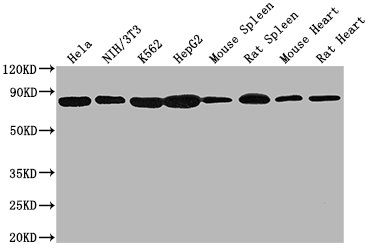
Western Blot Positive WB detected in: Hela whole cell lysate, NIH/3T3 whole cell lysate, K562 whole cell lysate, HepG2 whole cell lysate, Mouse spleen tissue, Rat spleen tissue, Mouse heart tissue, Rat heart tissue All lanes HSPA8 antibody at 1:2000 Secondary Goat polyclonal to mouse IgG at 1/50000 dilution Predicted band size: 70~75 KDa Observed band size: 70~75 KDa Exposure time: 10s |
|
|
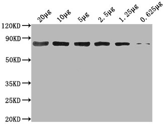
Western Blot Positive WB detected in: Hela whole cell lysate at 20ug, 10ug, 5ug, 2.5ug, 1.25ug, 0.625ugAll lanes: HSPA8 antibody at 1:2000 Secondary Goat polyclonal to mouse IgG at 1/50000 dilution Predicted band size: 70~75 KDa Observed band size: 70~75 KDa Exposure time: 10s |
|
|
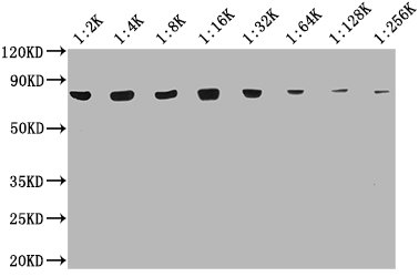
Western Blot Positive WB detected in: 20ug Hela whole cell lysate HSPA8 antibody at 1:2000, 1:4000, 1:8000, 1:16000, 1:32000, 1:64000, 1:128000, 1:256000 Secondary Goat polyclonal to mouse IgG at 1/50000 dilution Predicted band size: 70~75 KDa Observed band size: 70~75 KDa Exposure time: 10s |
Produktgarantie und fachkundiger Support
