
PRDX3 Monoclonal Antibody, IgG1, Clone: [6E6E12], Unconjugated, Mouse
Artikelnummer:
CSB-MA018656A1M
- Bilder (5)
| Artikelname: | PRDX3 Monoclonal Antibody, IgG1, Clone: [6E6E12], Unconjugated, Mouse |
| Artikelnummer: | CSB-MA018656A1M |
| Hersteller Artikelnummer: | CSB-MA018656A1m |
| Alternativnummer: | CSB-MA018656A1M-100UL, CSB-MA018656A1M-50UL |
| Hersteller: | Cusabio |
| Wirt: | Mouse |
| Kategorie: | Antikörper |
| Applikation: | ELISA, FC, IF, WB |
| Spezies Reaktivität: | Human |
| Konjugation: | Unconjugated |
| Alternative Synonym: | Antioxidant protein 1 antibody, AOP 1 antibody, AOP-1 antibody, AOP1 antibody, HBC189 antibody, MER5 antibody, MGC104387 antibody, MGC24293 antibody, mitochondrial antibody, peroxiredoxin 3 antibody, Peroxiredoxin III antibody, Peroxiredoxin-3 antibody, PRDX3 antibody, PRDX3_HUMAN antibody, PRO1748 antibody, Protein MER5 homolog antibody, PRX III antibody, Prx-III antibody, PRX3 antibody, SP 22 antibody, SP-22 antibody, SP22 antibody, Thioredoxin dependent peroxide reductase mitochondrial antibody, Thioredoxin-dependent peroxide reductase antibody |
| Klonalität: | Monoclonal |
| Klon-Bezeichnung: | [6E6E12] |
| Isotyp: | IgG1 |
| UniProt: | P30048 |
| Puffer: | Preservative: 0.03% Proclin 300<br />Constituents: 50% Glycerol, 0.01M PBS, PH 7.4 |
| Reinheit: | >95%, Protein G purified |
| Formulierung: | Liquid |
| Target-Kategorie: | PRDX3 |
| Application Verdünnung: | Recommended dilution: WB:1:1000-1:5000, IF:1:50-1:200, FC:1:50-1:200 |
|
|
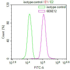
Overlay Peak curve showing Hela cells stained with CSB-MA018656A1m (red line) at 1:50. The cells were incubated in 10% normal goat serum to block non-specific protein-protein interactions followed by the antibody (1µg/1*106cells) for 1h at 4C. The secondary antibody used was FITC-conjugated Goat Anti-Mouse IgG(H+L) at 1/100 dilution for 30min at 4C. Isotype control antibody (green line) was mouse IgG1 (1µg/1*106cells) used under the same conditions. Acquisition of >10,000 events was performed. |
|
|
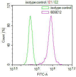
Overlay Peak curve showing MCF7 cells stained with CSB-MA018656A1m (red line) at 1:50. The cells were incubated in 10% normal goat serum to block non-specific protein-protein interactions followed by the antibody (1µg/1*106cells) for 1h at 4C. The secondary antibody used was FITC-conjugated Goat Anti-Mouse IgG(H+L) at 1/100 dilution for 30min at 4C. Isotype control antibody (green line) was mouse IgG1 (1µg/1*106cells) used under the same conditions. Acquisition of >10,000 events was performed. |
|
|
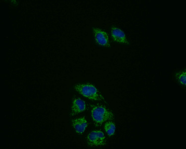
Immunofluorescence staining of Hela cells with(CSB-MA018656A1m)at 1:50, counter-stained with DAPI. The cells were fixed in 4% formaldehyde, permeabilized using 0.2% Triton X-100 and blocked in 10% normal Goat Serum. The cells were then incubated with the antibody overnight at 4C. Nuclear DNA was labeled in blue with DAPI. The secondary antibody was FITC-conjugated AffiniPure Goat Anti-Mouse IgG (H+L). |
|
|
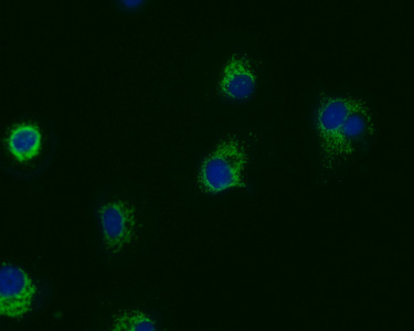
Immunofluorescence staining of MCF7 cells with(CSB-MA018656A1m)at 1:50, counter-stained with DAPI. The cells were fixed in 4% formaldehyde, permeabilized using 0.2% Triton X-100 and blocked in 10% normal Goat Serum. The cells were then incubated with the antibody overnight at 4C. Nuclear DNA was labeled in blue with DAPI. The secondary antibody was FITC-conjugated AffiniPure Goat Anti-Mouse IgG (H+L). |
|
|
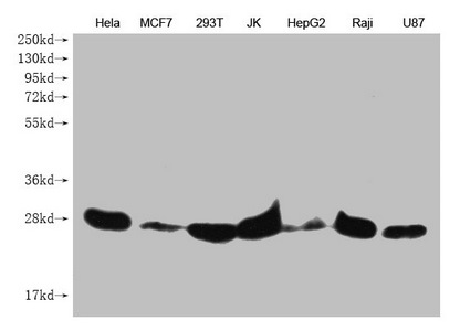
Western Blot Positive WB detected in: Hela whole cell lysate, MCF7 whole cell lysate, 293T whole cell lysate, JK whole cell lysate, HepG2 whole cell lysate, Raji whole cell lysate, U87 whole cell lysate All lanes: PRDX3 antibody at 1:1000 Secondary Goat polyclonal to mouse IgG at 1/50000 dilution Predicted band size: 28 kDa Observed band size: 28 KDa Exposure time:5min |
Produktgarantie und fachkundiger Support
