
SMURF1 Monoclonal Antibody, IgG2a, Clone: [1A9H4], Unconjugated, Mouse
Artikelnummer:
CSB-MA875720A0M
- Bilder (6)
| Artikelname: | SMURF1 Monoclonal Antibody, IgG2a, Clone: [1A9H4], Unconjugated, Mouse |
| Artikelnummer: | CSB-MA875720A0M |
| Hersteller Artikelnummer: | CSB-MA875720A0m |
| Alternativnummer: | CSB-MA875720A0M-100UL, CSB-MA875720A0M-50UL |
| Hersteller: | Cusabio |
| Wirt: | Mouse |
| Kategorie: | Antikörper |
| Applikation: | ELISA, FC, IF, IHC, WB |
| Spezies Reaktivität: | Human |
| Konjugation: | Unconjugated |
| Alternative Synonym: | E3 ubiquitin-protein ligase SMURF1 antibody, hSMURF1 antibody, KIAA1625 antibody, Smad specific E3 ubiquitin ligase 1 antibody, SMAD specific E3 ubiquitin protein ligase 1 antibody, Smad ubiquitination regulatory factor 1 antibody, SMAD-specific E3 ubiquitin-protein ligase 1 antibody, SMUF1_HUMAN antibody, SMURF 1 antibody, smurf1 antibody |
| Klonalität: | Monoclonal |
| Klon-Bezeichnung: | [1A9H4] |
| Isotyp: | IgG2a |
| UniProt: | Q9HCE7 |
| Puffer: | Preservative: 0.03% Proclin 300<br />Constituents: 50% Glycerol, 0.01M PBS, PH 7.4 |
| Reinheit: | >95%, Protein G purified |
| Formulierung: | Liquid |
| Target-Kategorie: | SMURF1 |
| Application Verdünnung: | Recommended dilution: WB:1:1000-1:5000, IHC:1:50-1:200, IF:1:50-1:200, FC:1:50-1:200 |
|
|
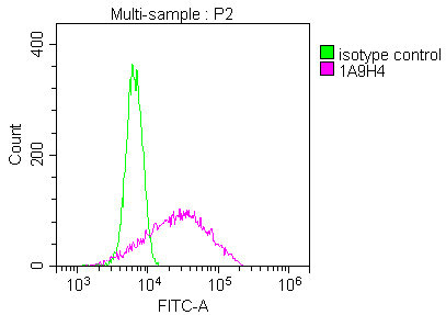
Overlay Peak curve showing Hela cells stained with CSB-MA875720A0m (red line) at 1:100. The cells were incubated in 10% normal goat serum to block non-specific protein-protein interactions followed by the antibody (1µg/1*106cells) for 1h at 4C. The secondary antibody used was FITC-conjugated Goat Anti-Mouse IgG(H+L) at 1/100 dilution for 30min at 4C. Isotype control antibody (green line) was mouse IgG1 (1µg/1*106cells) used under the same conditions. Acquisition of >10,000 events was performed. |
|
|
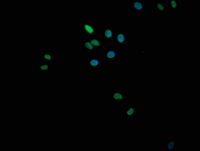
Immunofluorescence staining of Hela cells with CSB-MA875720A0m at 1:100, counter-stained with DAPI. The cells were fixed in 4% formaldehyde and blocked in 10% normal Goat Serum. The cells were incubated with the antibody overnight at 4C. Nuclear DNA was labeled in blue with DAPI. The secondary antibody was FITC-conjugated AffiniPure Goat Anti-Mouse IgG (H+L). |
|
|
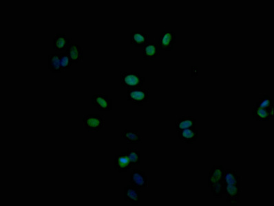
Immunofluorescence staining of HepG2 cells with CSB-MA875720A0m at 1:100, counter-stained with DAPI. The cells were fixed in 4% formaldehyde and blocked in 10% normal Goat Serum. The cells were incubated with the antibody overnight at 4C. Nuclear DNA was labeled in blue with DAPI. The secondary antibody was FITC-conjugated AffiniPure Goat Anti-Mouse IgG (H+L). |
|
|
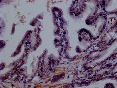
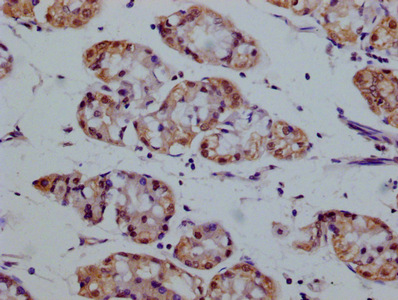
IHC image of CSB-MA875720A0m diluted at 1:100 and staining in paraffin-embedded human prostate tissue performed on a Leica BondTM system. After dewaxing and hydration, antigen retrieval was mediated by high pressure in a citrate buffer (pH 6.0). Section was blocked with 10% normal goat serum 30min at RT. Then primary antibody (1% BSA) was incubated at 4C overnight. The primary is detected by a biotinylated secondary antibody and visualized using an HRP conjugated SP system. |
|
|
IHC image of CSB-MA875720A0m diluted at 1:100 and staining in paraffin-embedded human stomach tissue performed on a Leica BondTM system. After dewaxing and hydration, antigen retrieval was mediated by high pressure in a citrate buffer (pH 6.0). Section was blocked with 10% normal goat serum 30min at RT. Then primary antibody (1% BSA) was incubated at 4C overnight. The primary is detected by a biotinylated secondary antibody and visualized using an HRP conjugated SP system. |
|
|
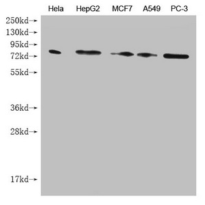
Western Blot Positive WB detected in: Hela whole cell lysate, HepG2 whole cell lysate, MCF7 whole cell lysate, A549 whole cell lysate, PC-3 whole cell lysate All lanes: SMURF1 antibody at 1:1000 Secondary Goat polyclonal to mouse IgG at 1/50000 dilution Predicted band size: 86 kDa Observed band size: 86 KDa Exposure time:5min |
Produktgarantie und fachkundiger Support
