
IFIT1 Antibody, Unconjugated, Rabbit, Polyclonal
Artikelnummer:
CSB-PA011018LA01HU
- Bilder (6)


| Artikelname: | IFIT1 Antibody, Unconjugated, Rabbit, Polyclonal |
| Artikelnummer: | CSB-PA011018LA01HU |
| Hersteller Artikelnummer: | CSB-PA011018LA01HU |
| Alternativnummer: | CSB-PA011018LA01HU-100UL |
| Hersteller: | Cusabio |
| Wirt: | Rabbit |
| Kategorie: | Antikörper |
| Applikation: | ELISA, IHC, WB |
| Spezies Reaktivität: | Human, Mouse, Rat |
| Konjugation: | Unconjugated |
| Alternative Synonym: | C56 antibody, G10P1 antibody, GARG 16 antibody, GARG16 antibody, IFI 56 antibody, IFI 56K antibody, IFI-56K antibody, IFI56 antibody, IFIT-1 antibody, Ifit1 antibody, IFIT1_HUMAN antibody, IFNAI1 antibody, Interferon alpha inducible protein (MW 56kD) antibody, Interferon induced 56 kDa protein antibody, Interferon induced protein 56 antibody, Interferon inducible mRNA 561 antibody, Interferon-induced 56 kDa protein antibody, Interferon-induced protein with tetratricopeptide repeats 1 antibody, ISG56 antibody, OTTHUMP00000020062 antibody, P56 antibody, RNM561 antibody |
| Klonalität: | Polyclonal |
| UniProt: | P09914 |
| Puffer: | PBS with 0.02% Sodium Azide, 50% Glycerol, pH 7.3. -20C, Avoid freeze / thaw cycles. |
| Reinheit: | Antigen Affinity Purified |
| Formulierung: | Liquid |
| Target-Kategorie: | IFIT1 |
| Application Verdünnung: | Recommended dilution: WB:1:1000-1:5000, IHC:1:20-1:200 |
|
|
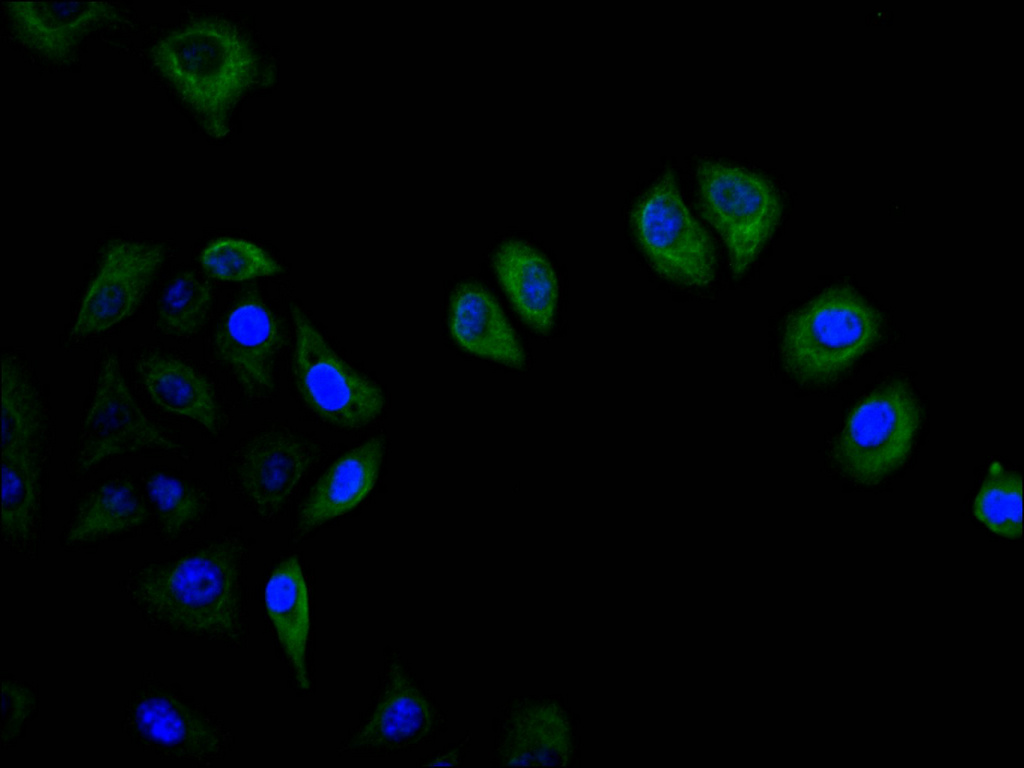
Immunofluorescent analysis of HepG2 |
|
|
IHC image of CSB-PA011018LA01HU diluted at 1:50 and staining in paraffin-embedded human adrenal gland tissue performed on a Leica BondTM system. After dewaxing and hydration, antigen retrieval was mediated by high pressure in a citrate buffer (pH 6.0). Section was blocked with 10% normal goat serum 30min at RT. Then primary antibody (1% BSA) was incubated at 4C overnight. The primary is detected by a Goat anti-rabbit polymer IgG labeled by HRP and visualized using 0.05% DAB.Secondary antibody only control: uses 1% BSA instead of primary antibody. |
|
|
IHC image of CSB-PA011018LA01HU diluted at 1:50 and staining in paraffin-embedded human liver cancer performed on a Leica BondTM system. After dewaxing and hydration, antigen retrieval was mediated by high pressure in a citrate buffer (pH 6.0). Section was blocked with 10% normal goat serum 30min at RT. Then primary antibody (1% BSA) was incubated at 4C overnight. The primary is detected by a Goat anti-rabbit polymer IgG labeled by HRP and visualized using 0.05% DAB. Secondaryantibodyonlycontrol: uses 1%BSA instead of primary antibody. |
|
|
IHC image of CSB-PA011018LA01HU diluted at 1:50 and staining in paraffin-embedded human breast cancer performed on a Leica BondTM system. After dewaxing and hydration, antigen retrieval was mediated by high pressure in a citrate buffer (pH 6.0). Section was blocked with 10% normal goat serum 30min at RT. Then primary antibody (1% BSA) was incubated at 4C overnight. The primary is detected by a Goat anti-rabbit polymer IgG labeled by HRP and visualized using 0.05% DAB.Secondaryantibodyonlycontrol: uses 1%BSA instead of primary antibody. |
|
|
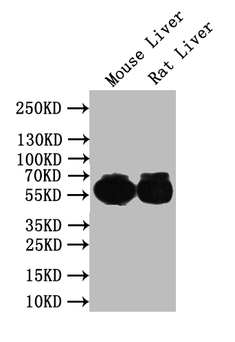
Western BlotPositive WB detected in: Mouse Liver tissue lysate, Rat Liver tissue lysateAll lanes: IFIT1 antibody at 1:1000SecondaryGoat polyclonal to rabbit IgG at 1/50000 dilutionPredicted band size: 56 kDaObserved band size: 56 kDa |
|
|
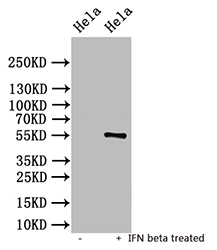
Western Blot Positive WB detected in: untreated Hela whole cell lysate, IFN beta treated Hela whole cell lysate All lanes: IFIT1 antibody at 1:1000 Secondary Goat polyclonal to rabbit IgG at 1/50000 dilution Predicted band size: 56 kDa Observed band size: 56 kDa |
Produktgarantie und fachkundiger Support
