
LANCL1 Antibody, Unconjugated, Rabbit, Polyclonal
Artikelnummer:
CSB-PA012742LA01HU
- Bilder (5)


| Artikelname: | LANCL1 Antibody, Unconjugated, Rabbit, Polyclonal |
| Artikelnummer: | CSB-PA012742LA01HU |
| Hersteller Artikelnummer: | CSB-PA012742LA01HU |
| Alternativnummer: | CSB-PA012742LA01HU-100UG, CSB-PA012742LA01HU-50UG |
| Hersteller: | Cusabio |
| Wirt: | Rabbit |
| Kategorie: | Antikörper |
| Applikation: | ELISA, IHC, WB |
| Spezies Reaktivität: | Human, Mouse, Rat |
| Konjugation: | Unconjugated |
| Alternative Synonym: | 40 kDa erythrocyte membrane protein antibody, G protein-coupled receptor 69A antibody, GPR69A antibody, LanC (bacterial lantibiotic synthetase component C) like 1 antibody, LanC lantibiotic synthetase component C like 1 (bacterial) antibody, LanC-like protein 1 antibody, LANC1_HUMAN antibody, LANCL1 antibody, p40 antibody |
| Klonalität: | Polyclonal |
| UniProt: | O43813 |
| Puffer: | Preservative: Liquid in PBS containing 50% glycerol, 0.5% BSA and 0.02% sodium azide. |
| Reinheit: | Antigen Affinity Purified |
| Formulierung: | Liquid |
| Target-Kategorie: | LANCL1 |
| Application Verdünnung: | Recommended dilution: WB:1:1000-1:5000 , IHC:1:200-1:500 |
|
|
|
|
|
|
|
|
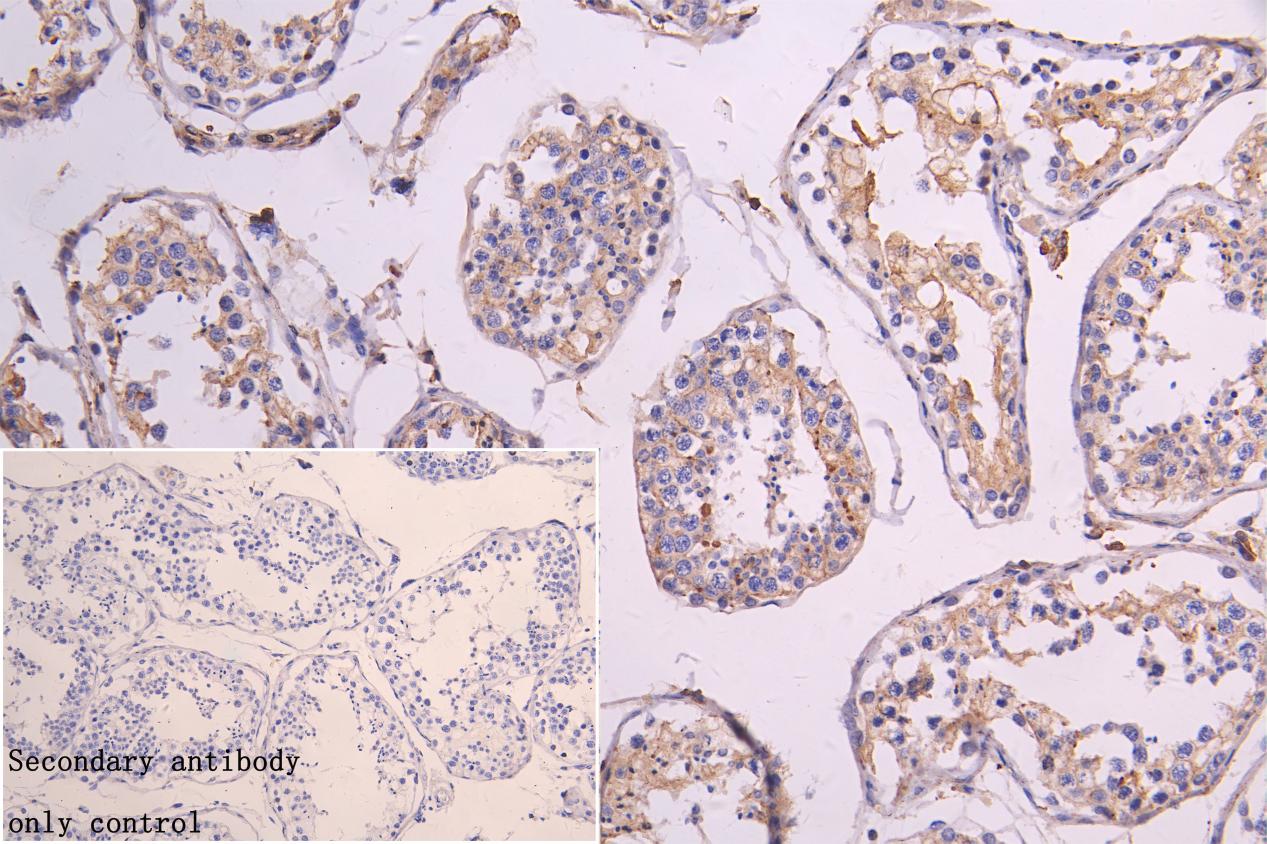
IHC image of CSB-PA012742LA01HU diluted at 1:300 and staining in paraffin-embedded human testis tissue performed on a Leica BondTM system. After dewaxing and hydration, antigen retrieval was mediated by high pressure in a citrate buffer (pH 6.0). Section was blocked with 10% normal goat serum 30min at RT. Then primary antibody (1% BSA) was incubated at 4C overnight. The primary is detected by a Goat anti-rabbit polymer IgG labeled by HRP and visualized using 0.05% DAB.Secondary antibody only control: uses 1% BSA instead of primary antibody |
|
|
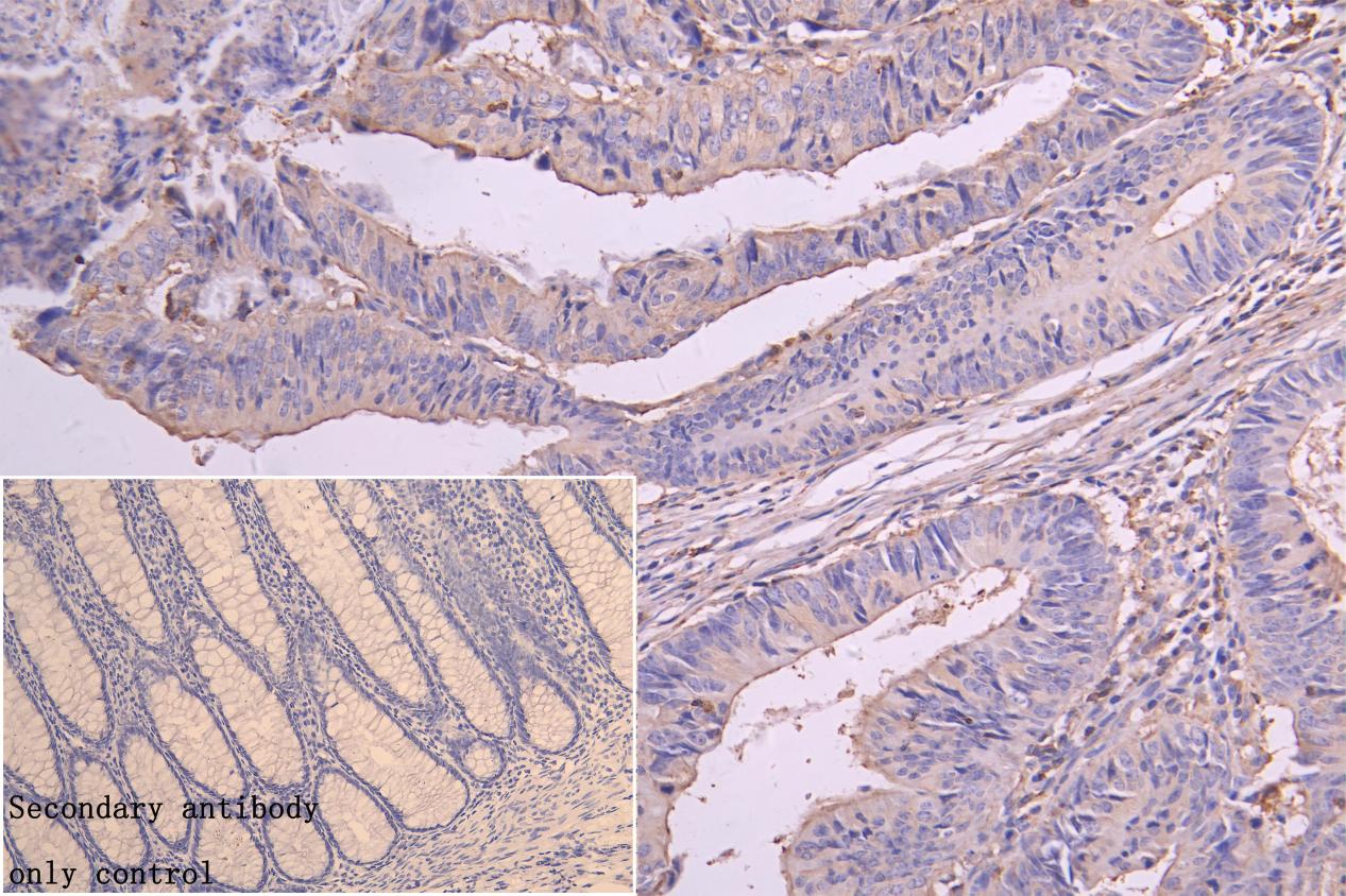
IHC image of CSB-PA012742LA01HU diluted at 1:300 and staining in paraffin-embedded human colorectal cancer performed on a Leica BondTM system. After dewaxing and hydration, antigen retrieval was mediated by high pressure in a citrate buffer (pH 6.0). Section was blocked with 10% normal goat serum 30min at RT. Then primary antibody (1% BSA) was incubated at 4C overnight. The primary is detected by a Goat anti-rabbit polymer IgG labeled by HRP and visualized using 0.05% DAB.Secondary antibody only control: uses 1% BSA instead of primary antibody |
|
|
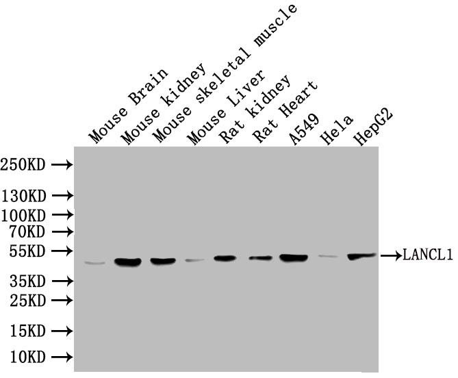
Western Blot Positive WB detected in: Mouse Brain tissue lysate, Mouse Kidney tissue lysate, Mouse skeletal muscle tissue lysate, Mous Liver tissue lysate, Rat Kidney tissue lysate, Rat Heart tissue lysate, A549 whole cell lysate, Hela whole cell lysate, HepG2 whole cell lysate All lanes: LANCL1 antibody at 1:1000 Secondary Goat polyclonal to rabbit IgG at 1/50000 dilution Predicted band size: 42 kDa Observed band size: 42 kDa |
Produktgarantie und fachkundiger Support
