
PRKN Antibody, Unconjugated, Rabbit, Polyclonal
Artikelnummer:
CSB-PA017451LA01HU
- Bilder (4)
| Artikelname: | PRKN Antibody, Unconjugated, Rabbit, Polyclonal |
| Artikelnummer: | CSB-PA017451LA01HU |
| Hersteller Artikelnummer: | CSB-PA017451LA01HU |
| Alternativnummer: | CSB-PA017451LA01HU-100UG, CSB-PA017451LA01HU-50UG |
| Hersteller: | Cusabio |
| Wirt: | Rabbit |
| Kategorie: | Antikörper |
| Applikation: | ELISA, IF, IHC, WB |
| Spezies Reaktivität: | Human |
| Konjugation: | Unconjugated |
| Alternative Synonym: | AR JP antibody, E3 ubiquitin ligase antibody, E3 ubiquitin protein ligase parkin antibody, E3 ubiquitin-protein ligase parkin antibody, FRA6E antibody, LPRS 2 antibody, LPRS2 antibody, PARK 2 antibody, Park2 antibody, Parkin 2 antibody, Parkinson disease (autosomal recessive juvenile) 2 antibody, Parkinson disease (autosomal recessive, juvenile) 2, parkin antibody, Parkinson disease protein 2 antibody, Parkinson juvenile disease protein 2 antibody, Parkinson protein 2 E3 ubiquitin protein ligase antibody, Parkinson protein 2, E3 ubiquitin protein ligase (parkin) antibody, PDJ antibody, PRKN 2 antibody, PRKN antibody, PRKN2 antibody, PRKN2_HUMAN antibody, Ubiquitin E3 ligase PRKN antibody |
| Klonalität: | Polyclonal |
| UniProt: | O60260 |
| Puffer: | Preservative: 0.03% Proclin 300<br />Constituents: 50% Glycerol, 0.01M PBS, PH 7.4 |
| Reinheit: | >95%, Protein G purified |
| Formulierung: | Liquid |
| Target-Kategorie: | PRKN |
| Application Verdünnung: | Recommended dilution: WB:1:500-1:5000, IHC:1:500-1:1000, IF:1:50-1:500 |
|
|
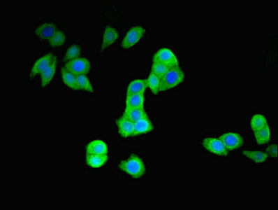
Immunofluorescent analysis of PC-3 cells using CSB-PA017451LA01HU at dilution of 1:100 and Alexa Fluor 488-congugated AffiniPure Goat Anti-Rabbit IgG(H+L) |
|
|
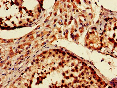
IHC image of CSB-PA017451LA01HU diluted at 1:600 and staining in paraffin-embedded human testis tissue performed on a Leica BondTM system. After dewaxing and hydration, antigen retrieval was mediated by high pressure in a citrate buffer (pH 6.0). Section was blocked with 10% normal goat serum 30min at RT. Then primary antibody (1% BSA) was incubated at 4°,C overnight. The primary is detected by a biotinylated secondary antibody and visualized using an HRP conjugated SP system. |
|
|
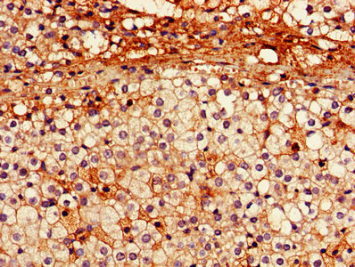
IHC image of CSB-PA017451LA01HU diluted at 1:600 and staining in paraffin-embedded human adrenal gland tissue performed on a Leica BondTM system. After dewaxing and hydration, antigen retrieval was mediated by high pressure in a citrate buffer (pH 6.0). Section was blocked with 10% normal goat serum 30min at RT. Then primary antibody (1% BSA) was incubated at 4°,C overnight. The primary is detected by a biotinylated secondary antibody and visualized using an HRP conjugated SP system. |
|
|
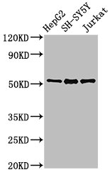
Western Blot Positive WB detected in: HepG2 whole cell lysate, SH-SY5Y whole cell lysate, Jurkat whole cell lysate All lanes: PRKN antibody at 3µg/ml Secondary Goat polyclonal to rabbit IgG at 1/50000 dilution Predicted band size: 52, 49, 24, 31, 43, 36, 44, 47 kDa Observed band size: 52 kDa |
Produktgarantie und fachkundiger Support
