
PTBP3 Antibody, Unconjugated, Rabbit, Polyclonal
Artikelnummer:
CSB-PA020060NA01HU
- Bilder (9)


| Artikelname: | PTBP3 Antibody, Unconjugated, Rabbit, Polyclonal |
| Artikelnummer: | CSB-PA020060NA01HU |
| Hersteller Artikelnummer: | CSB-PA020060NA01HU |
| Alternativnummer: | CSB-PA020060NA01HU-100UL, CSB-PA020060NA01HU-50UL |
| Hersteller: | Cusabio |
| Wirt: | Rabbit |
| Kategorie: | Antikörper |
| Applikation: | ELISA, IHC, WB |
| Spezies Reaktivität: | Human |
| Konjugation: | Unconjugated |
| Alternative Synonym: | DKFZp781I1117 antibody, Fission yeast differentiation regulator antibody, OTTHUMP00000021931 antibody, OTTHUMP00000021932 antibody, Polypyrimidine tract binding protein 3 antibody, regulator of differentiation 1 antibody, Regulator of differentiation (in S. pombe) 1 antibody, Regulator of differentiation 1 antibody, ROD 1 antibody, Rod1 antibody, ROD1 regulator of differentiation 1 antibody, ROD1_HUMAN antibody |
| Klonalität: | Polyclonal |
| UniProt: | O95758 |
| Puffer: | pH7.4 PBS, 0.05% NaN3, 40% Glycerol |
| Reinheit: | Antigen Affinity Purified |
| Formulierung: | Liquid |
| Target-Kategorie: | PTBP3 |
| Application Verdünnung: | Recommended dilution: WB:1:1000-1:5000, IHC:1:20-1:200 |
|
|
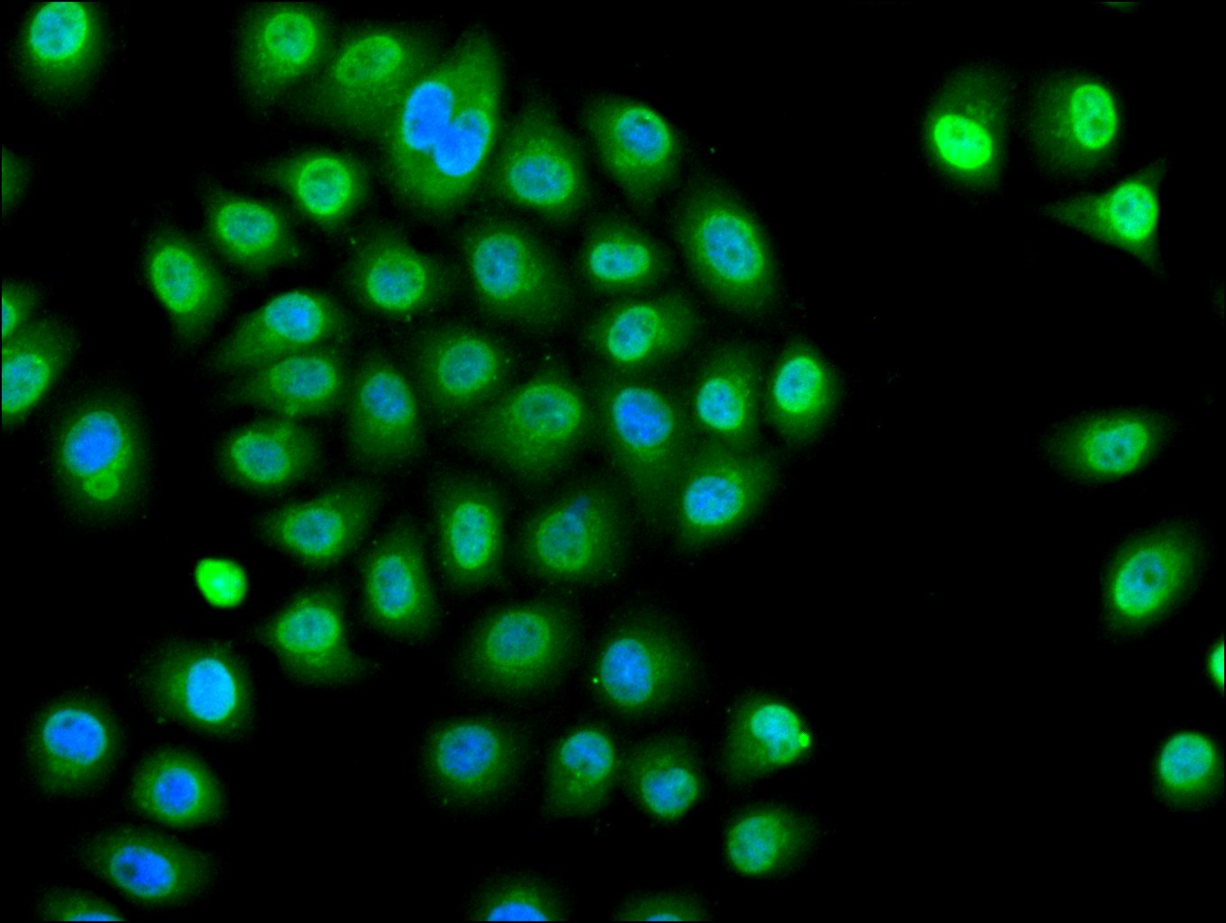
Immunofluorescence staining of A549 cell with CSB-PA020060NA01HU at 1:30, counter-stained with DAPI. The cells were fixed in 4% formaldehyde and blocked in 10% normal Goat Serum. The cells were then incubated with the antibody overnight at 4C. The secondary antibody was Alexa Fluor 488-congugated AffiniPure Goat Anti-Rabbit IgG(H+L). |
|
|
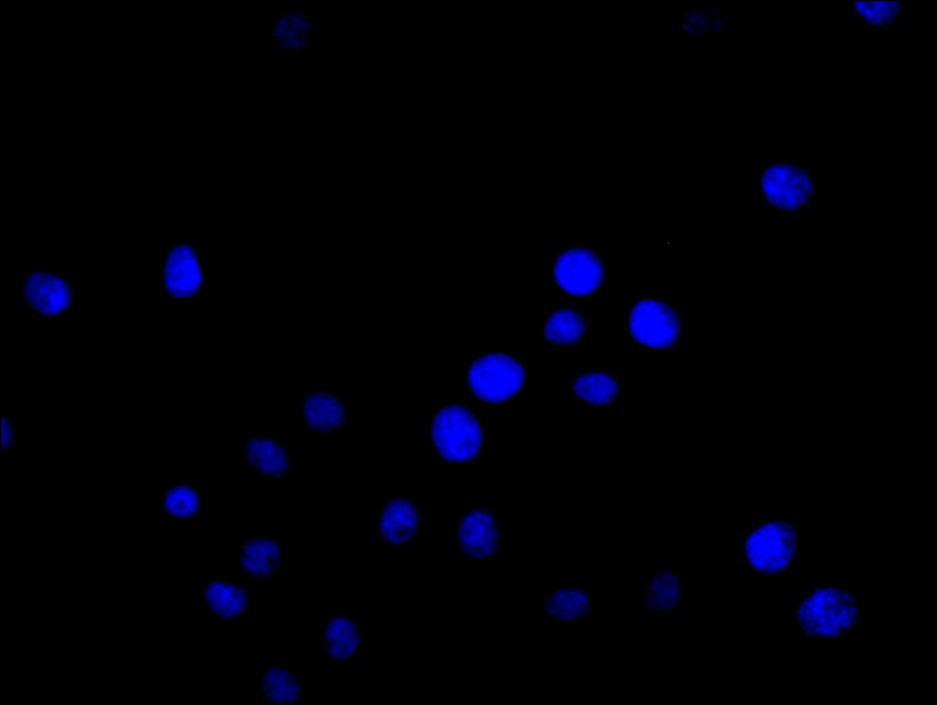
Immunofluorescence staining of A549 cell with 5% goat serum, counter-stained with DAPI. The cells were fixed in 4% formaldehyde and blocked in 10% normal Goat Serum. The cells were then incubated with the antibody overnight at 4C. The secondary antibody was Alexa Fluor 488-congugated AffiniPure Goat Anti-Rabbit IgG(H+L). |
|
|
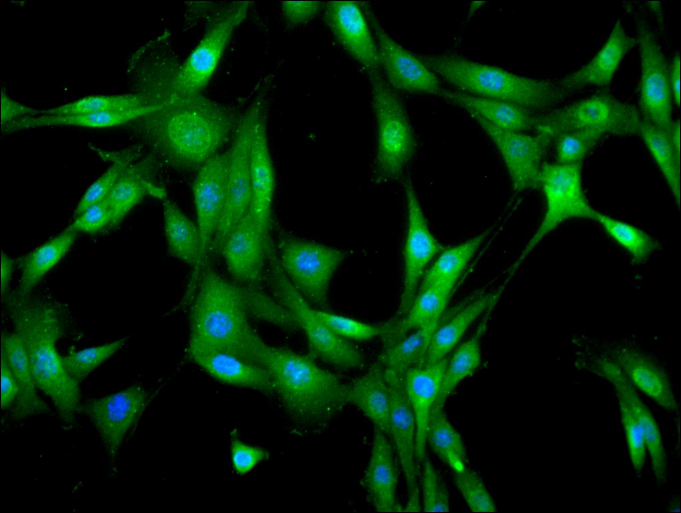
Immunofluorescence staining of NIH/3T3 cell with CSB-PA020060NA01HU at 1:30, counter-stained with DAPI. The cells were fixed in 4% formaldehyde and blocked in 10% normal Goat Serum. The cells were then incubated with the antibody overnight at 4C. The secondary antibody was Alexa Fluor 488-congugated AffiniPure Goat Anti-Rabbit IgG(H+L). |
|
|
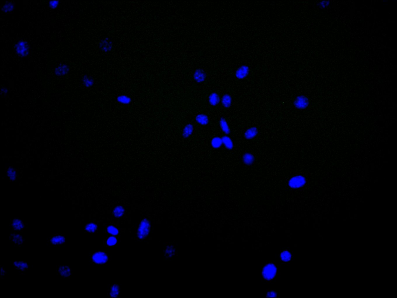
Immunofluorescence staining of NIH/3T3 cell with 5% goat serum, counter-stained with DAPI. The cells were fixed in 4% formaldehyde and blocked in 10% normal Goat Serum. The cells were then incubated with the antibody overnight at 4C. The secondary antibody was Alexa Fluor 488-congugated AffiniPure Goat Anti-Rabbit IgG(H+L). |
|
|
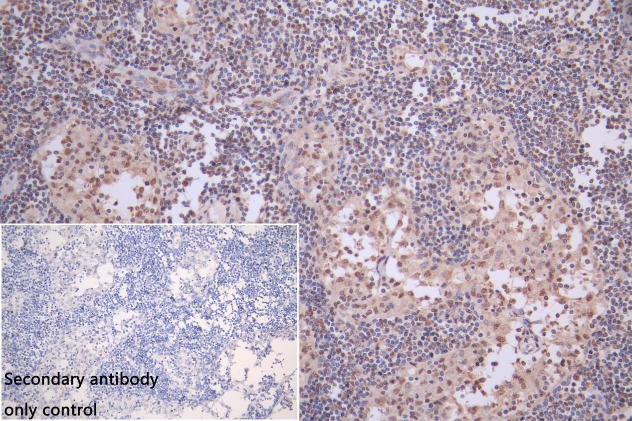
IHC image of CSB-PA020060NA01HU diluted at 1:200 and staining in paraffin-embedded human Lymphnode tissue performed on a Leica BondTM system. After dewaxing and hydration, antigen retrieval was mediated by high pressure in a citrate buffer (pH 6.0). Section was blocked with 10% normal goat serum 30min at RT. Then primary antibody (1% BSA) was incubated at 4C overnight. The primary is detected by a Goat anti-rabbit polymer IgG labeled by HRP and visualized using 0.05% DAB. |
|
|
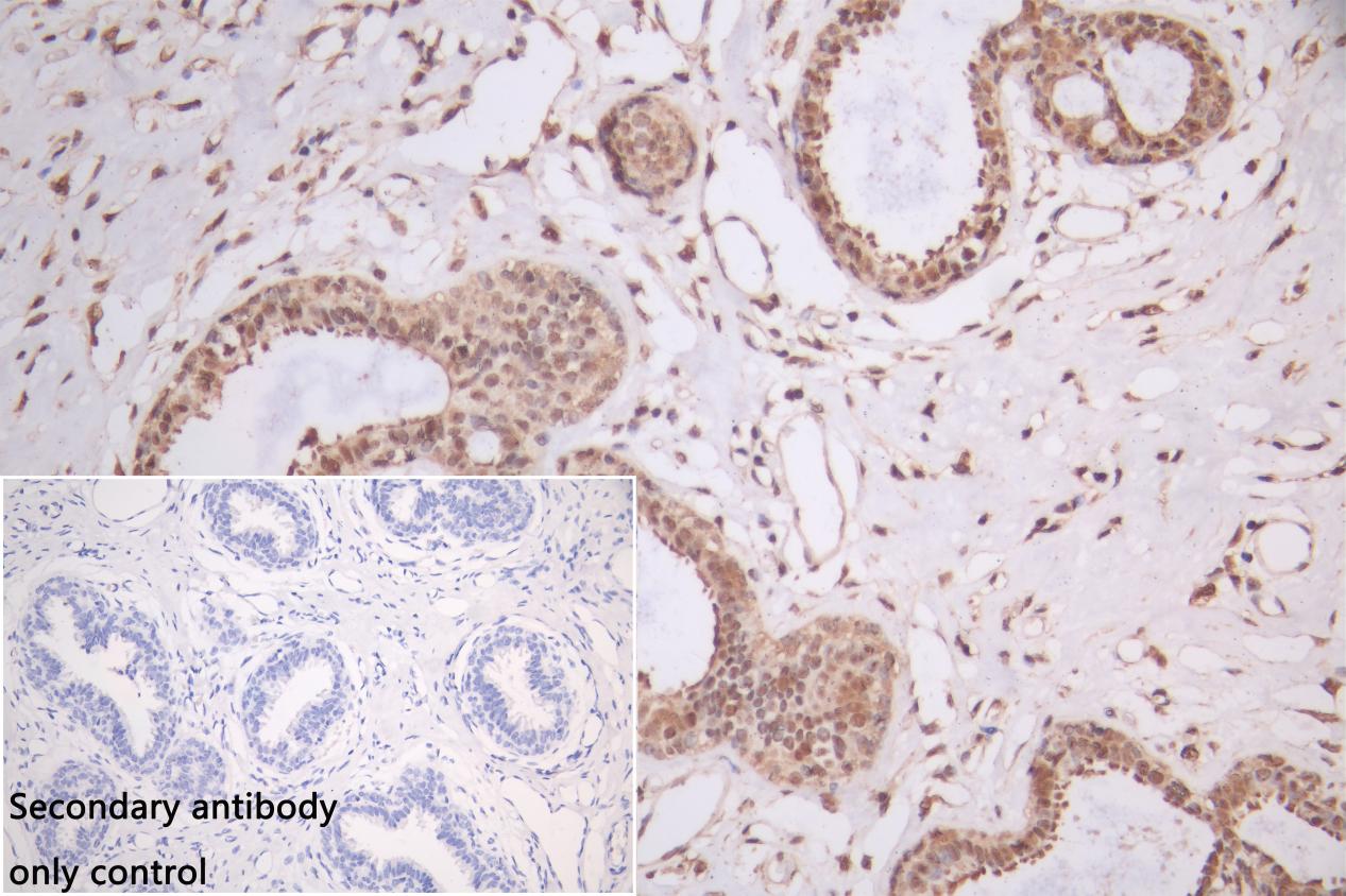
IHC image of CSB-PA020060NA01HU diluted at 1:200 and staining in paraffin-embedded human breast cancer performed on a Leica BondTM system. After dewaxing and hydration, antigen retrieval was mediated by high pressure in a citrate buffer (pH 6.0). Section was blocked with 10% normal goat serum 30min at RT. Then primary antibody (1% BSA) was incubated at 4C overnight. The primary is detected by a Goat anti-rabbit polymer IgG labeled by HRP and visualized using 0.05% DAB.Secondary antibody only control: uses 1% BSA instead of primary antibody |
|
|
|
|
|
Western Blot Positive WB detected in: K562 whole cell lysate, MCF-7 whole cell lysate All lanes: PTBP3 antibody at 1:2000 Secondary Goat polyclonal to rabbit IgG at 1/50000 dilution Predicted band size: 60, 57, 61, 50 kDa Observed band size: 60 kDa |
|
|
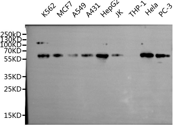
Western Blot Positive WB detected in: K562 whole cell lysate, MCF7 whole cell lysate, A549 whole cell lysate, A431 whole cell lysate, HepG2 whole cell lysate, JK whole cell lysate, Hela whole cell lysate,PC-3 whole cell lysate All lanes: PTBP3 antibody at 1:1000 Secondary Goat polyclonal to rabbit IgG at 1/50000 dilution Predicted band size: 55,60kDa Observed band size: 55 kDa |
Produktgarantie und fachkundiger Support
