
RPL19 Antibody, Unconjugated, Rabbit, Polyclonal
Artikelnummer:
CSB-PA020160LA01HU
- Bilder (6)


| Artikelname: | RPL19 Antibody, Unconjugated, Rabbit, Polyclonal |
| Artikelnummer: | CSB-PA020160LA01HU |
| Hersteller Artikelnummer: | CSB-PA020160LA01HU |
| Alternativnummer: | CSB-PA020160LA01HU-100UL |
| Hersteller: | Cusabio |
| Wirt: | Rabbit |
| Kategorie: | Antikörper |
| Applikation: | ELISA, IF, IHC, WB |
| Spezies Reaktivität: | Human, Mouse |
| Konjugation: | Unconjugated |
| Alternative Synonym: | 60S ribosomal protein L19 antibody, DKFZp779D216 antibody, FLJ27452 antibody, HGNC:10312 antibody, L19 antibody, MGC71997 antibody, Ribosomal Protein L19 antibody, Ribosomal protein L19, cytosolic, N terminus truncated antibody, RL19_HUMAN antibody, rpl19 antibody |
| Klonalität: | Polyclonal |
| UniProt: | P84098 |
| Puffer: | Preservative: 0.03% Proclin 300<br /> Constituents: 50% Glycerol, 0.01M PBS, PH 7.4 |
| Reinheit: | Antigen Affinity Purified |
| Formulierung: | Liquid |
| Target-Kategorie: | RPL19 |
| Application Verdünnung: | Recommended dilution: WB:1:500-1:2000,IHC:1:20-1:200,IF:1:20-1:200 |
|
|
Imm |
|
|
|
|
|
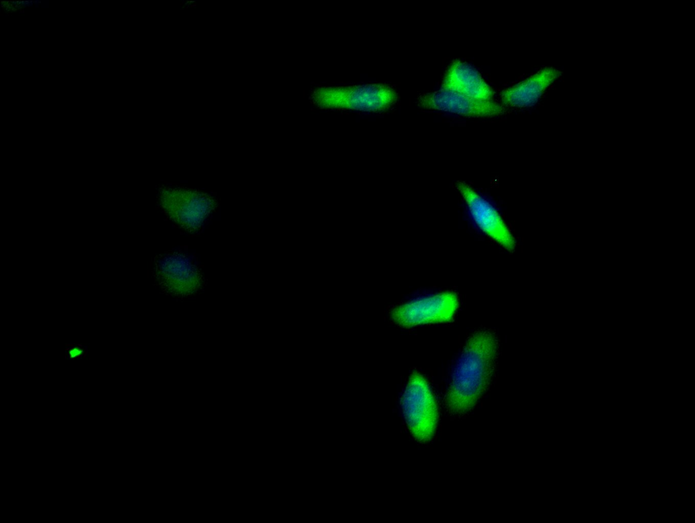
Immunofluorescence staining of HepG2 cell with CSB-PA020160LA01HU at 1:25, counter-stained with DAPI. The cells were fixed in 4% formaldehyde and and permeated by 0.2% TritonX-100 for 15 min. Then 10% normal goat serum to block non-specific protein-protein interactions . The cells were then incubated with the antibody overnight at 4°C. The secondary antibody was Alexa Fluor 488-congugated AffiniPure Goat Anti-Rabbit IgG(H+L). |
|
|
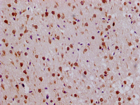
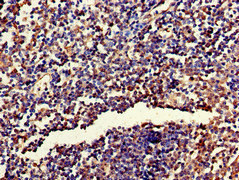
IHC image of CSB-PA020160LA01HU diluted at 1:50 and staining in paraffin-embedded human endometrial cancer performed on a Leica BondTM system. After dewaxing and hydration, antigen retrieval was mediated by high pressure in a citrate buffer (pH 6.0). Section was blocked with 10% normal goat serum 30min at RT. Then primary antibody (1% BSA) was incubated at 4C overnight. The primary is detected by a Goat anti-rabbit polymer IgG labeled by HRP and visualized using 0.05% DAB. |
|
|
IHC image of CSB-PA020160LA01HU diluted at 1:50 and staining in paraffin-embedded human prostate tissue performed on a Leica BondTM system. After dewaxing and hydration, antigen retrieval was mediated by high pressure in a citrate buffer (pH 6.0). Section was blocked with 10% normal goat serum 30min at RT. Then primary antibody (1% BSA) was incubated at 4C overnight. The primary is detected by a Goat anti-rabbit polymer IgG labeled by HRP and visualized using 0.05% DAB. |
|
|
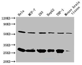
Western Blot Positive WB detected in: Hela whole cell lysate(20µg), MCF7 whole cell lysate(20µg), PC-3 whole cell lysate(20µg), HEK293 whole cell lysate(20µg), HepG2 whole cell lysate(20µg) All lanes: RPL19 antibody at 1:500 Secondary Goat polyclonal to rabbit IgG at 1/40000 dilution Predicted band size: 23,28 kDa Observed band size: 28 kDa Exposure time: 180s |
Produktgarantie und fachkundiger Support
