
SPCS3 Antibody, Unconjugated, Rabbit, Polyclonal
Artikelnummer:
CSB-PA022517LA01HU
- Bilder (6)


| Artikelname: | SPCS3 Antibody, Unconjugated, Rabbit, Polyclonal |
| Artikelnummer: | CSB-PA022517LA01HU |
| Hersteller Artikelnummer: | CSB-PA022517LA01HU |
| Alternativnummer: | CSB-PA022517LA01HU-100UL |
| Hersteller: | Cusabio |
| Wirt: | Rabbit |
| Kategorie: | Antikörper |
| Applikation: | ELISA, IHC, WB |
| Spezies Reaktivität: | Human, Mouse |
| Konjugation: | Unconjugated |
| Alternative Synonym: | SPCS3 antibody, SPC22 antibody, UNQ1841/PRO3567 antibody, Signal peptidase complex subunit 3 antibody, EC 3.4.-.- antibody, Microsomal signal peptidase 22/23 kDa subunit antibody, SPC22/23 antibody, SPase 22/23 kDa subunit antibody |
| Klonalität: | Polyclonal |
| UniProt: | P61009 |
| Puffer: | Preservative: PBS with 0.02% sodium azide and 50% glycerol pH 7.3 |
| Reinheit: | Antigen Affinity Purified |
| Formulierung: | Liquid |
| Target-Kategorie: | SPCS3 |
| Application Verdünnung: | Recommended dilution: WB:1:500-1:2000, IHC:1:20-1:200 |
|
|
Immunohistochemistry of paraffin-embedded human liver tissue using CSB-PA022517LA01HU at dilution of 1:100 |
|
|
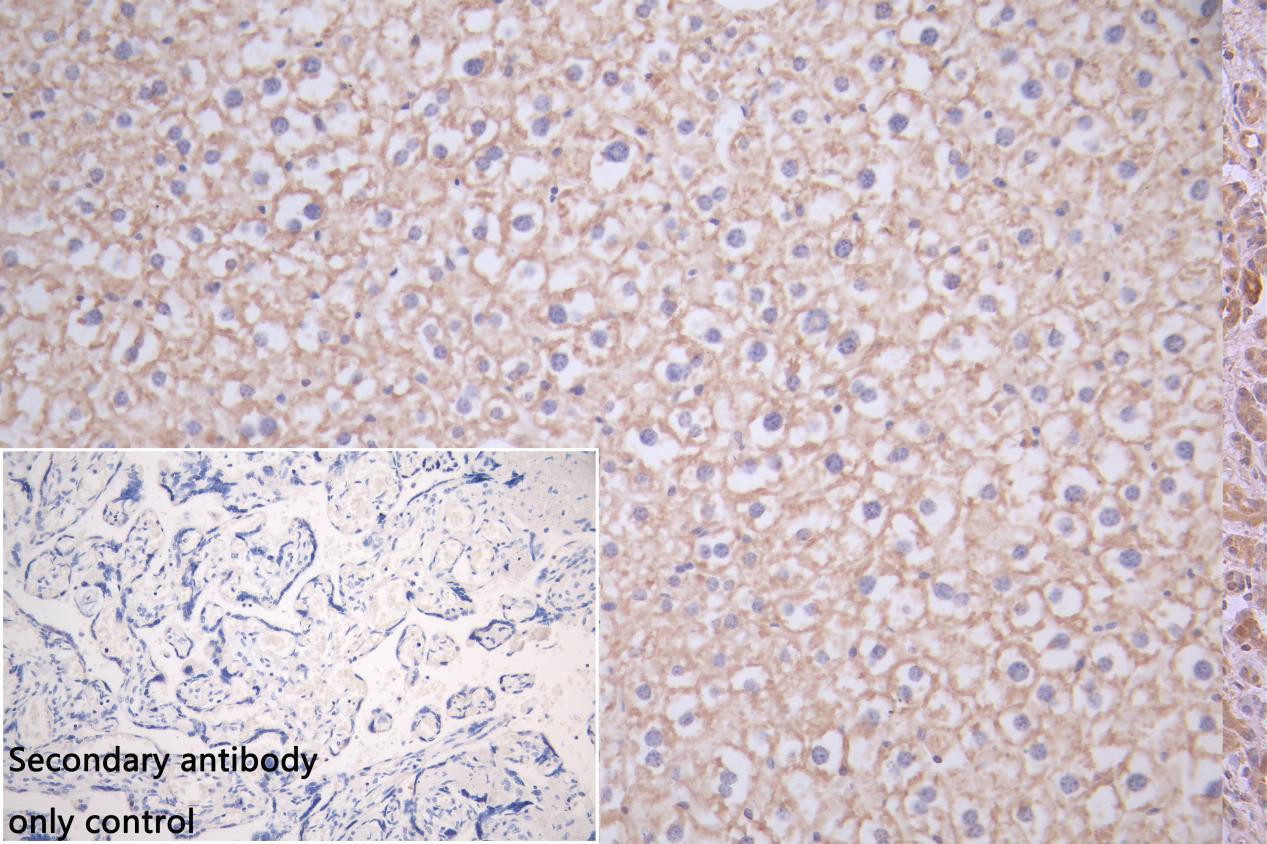
IHC image of CSB-PA022517LA01HU diluted at 1:150 and staining in paraffin-embedded mouse liver tissue performed on a Leica BondTM system. After dewaxing and hydration, antigen retrieval was mediated by high pressure in a citrate buffer (pH 6.0). Section was blocked with 10% normal goat serum 30min at RT. Then primary antibody (1% BSA) was incubated at 4C overnight. The primary is detected by a Goat anti-rabbit polymer IgG labeled by HRP and visualized using 0.05% DAB.Secondary antibody only control: uses 1% BSA instead of primary antibody |
|
|
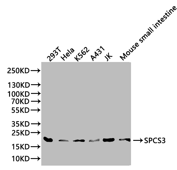
Western Blot Positive WB detected in: 293T whole cell lysate, Hela whole cell lysate, K562 whole cell lysate, A431 whole cell lysate, JK whole cell lysate, Mouset small intestinetissue lysate All lanes: SPCS3 antibody at 1:1000 Secondary Goat polyclonal to rabbit IgG at 1/50000 dilution Predicted band size: 21 kDa Observed band size: 21kDa |
|
|
Immunohistochemistry of paraffin-embedded human placenta tissue using CSB-PA022517LA01HU at dilution of 1:100 |
|
|
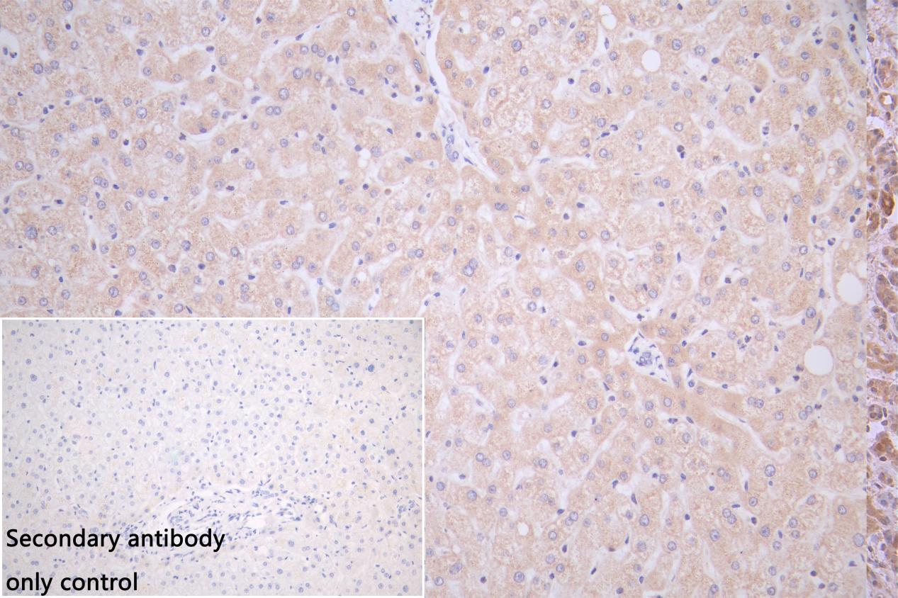
IHC image of CSB-PA022517LA01HU diluted at 1:150 and staining in paraffin-embedded human liver tissue performed on a Leica BondTM system. After dewaxing and hydration, antigen retrieval was mediated by high pressure in a citrate buffer (pH 6.0). Section was blocked with 10% normal goat serum 30min at RT. Then primary antibody (1% BSA) was incubated at 4C overnight. The primary is detected by a Goat anti-rabbit polymer IgG labeled by HRP and visualized using 0.05% DAB. |
|
|
IHC image of CSB-PA022517LA01HU diluted at 1:150 and staining in paraffin-embedded human placenta tissue performed on a Leica BondTM system. After dewaxing and hydration, antigen retrieval was mediated by high pressure in a citrate buffer (pH 6.0). Section was blocked with 10% normal goat serum 30min at RT. Then primary antibody (1% BSA) was incubated at 4C overnight. The primary is detected by a Goat anti-rabbit polymer IgG labeled by HRP and visualized using 0.05% DAB.Secondary antibody only control: uses 1% BSA instead of primary antibody |
Produktgarantie und fachkundiger Support
