
TP53 Antibody, Unconjugated, Rabbit, Polyclonal
Artikelnummer:
CSB-PA07889A0RB
- Bilder (9)


| Artikelname: | TP53 Antibody, Unconjugated, Rabbit, Polyclonal |
| Artikelnummer: | CSB-PA07889A0RB |
| Hersteller Artikelnummer: | CSB-PA07889A0Rb |
| Alternativnummer: | CSB-PA07889A0RB-100UG, CSB-PA07889A0RB-50UG |
| Hersteller: | Cusabio |
| Wirt: | Rabbit |
| Kategorie: | Antikörper |
| Applikation: | ELISA, IF, WB |
| Spezies Reaktivität: | Human, Rat |
| Konjugation: | Unconjugated |
| Alternative Synonym: | Antigen NY-CO-13 antibody, BCC7 antibody, Cellular tumor antigen p53 antibody, FLJ92943 antibody, LFS1 antibody, Mutant tumor protein 53 antibody, p53 antibody, p53 tumor suppressor antibody, P53_HUMAN antibody, Phosphoprotein p53 antibody, Tp53 antibody, Transformation related protein 53 antibody, TRP53 antibody, tumor antigen p55 antibody, Tumor protein 53 antibody, Tumor protein p53 antibody, Tumor suppressor p53 antibody |
| Klonalität: | Polyclonal |
| UniProt: | P04637 |
| Puffer: | Preservative: 0.03% Proclin 300<br />Constituents: 50% Glycerol, 0.01M PBS, PH 7.4 |
| Reinheit: | >95%, Protein G purified |
| Formulierung: | Liquid |
| Target-Kategorie: | TP53 |
| Application Verdünnung: | Recommended dilution: WB:1:500-1:1000,IF:1:20-1:100 |
|
|
|
|
|
|
|
|
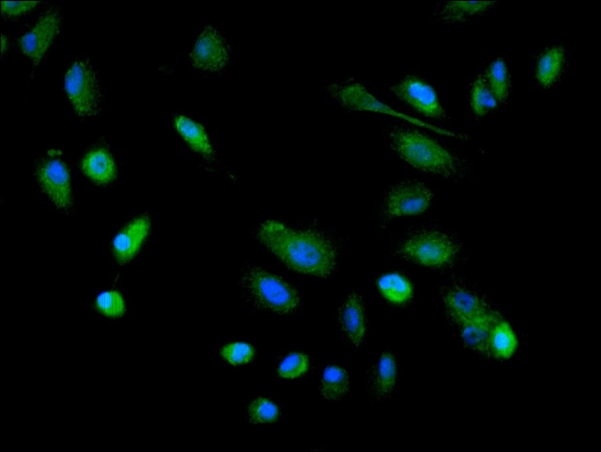
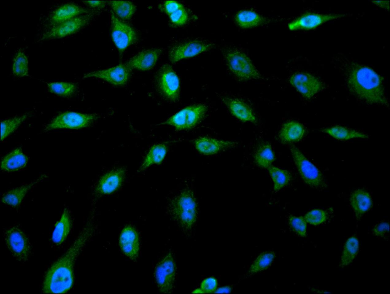
Immunofluorescence staining of A549 cell with CSB-PA07889A0Rb at 1:30, counter-stained with DAPI. The cells were fixed in 4% formaldehyde and blocked in 10% normal Goat Serum. The cells were then incubated with the antibody overnight at 4C. The secondary antibody was Alexa Fluor 488-congugated AffiniPure Goat Anti-Rabbit IgG(H+L). |
|
|
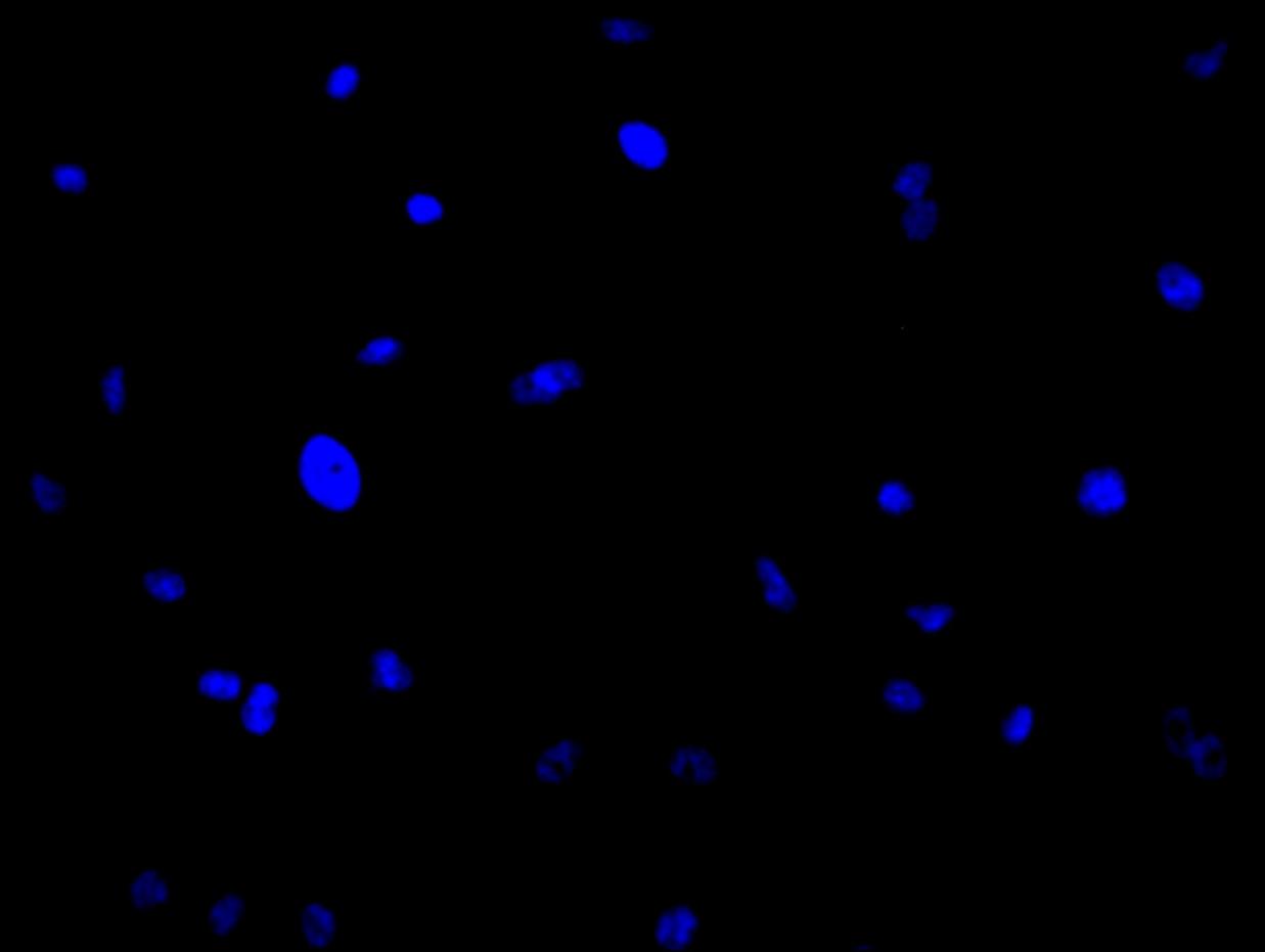
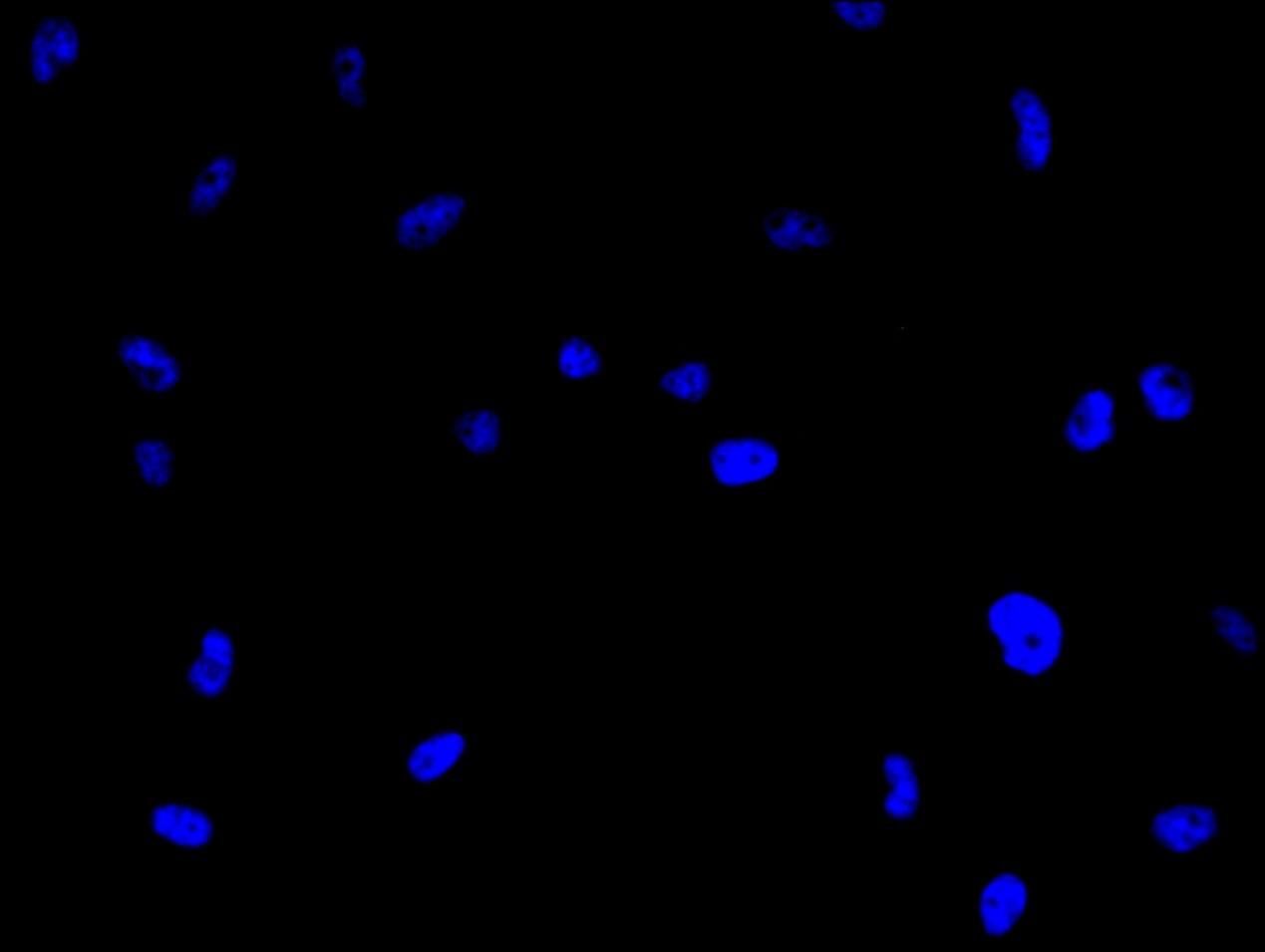
Immunofluorescence staining of A549 cell with 5% goat serum, counter-stained with DAPI. The cells were fixed in 4% formaldehyde and blocked in 10% normal Goat Serum. The cells were then incubated with the antibody overnight at 4C. The secondary antibody was Alexa Fluor 488-congugated AffiniPure Goat Anti-Rabbit IgG(H+L). |
|
|
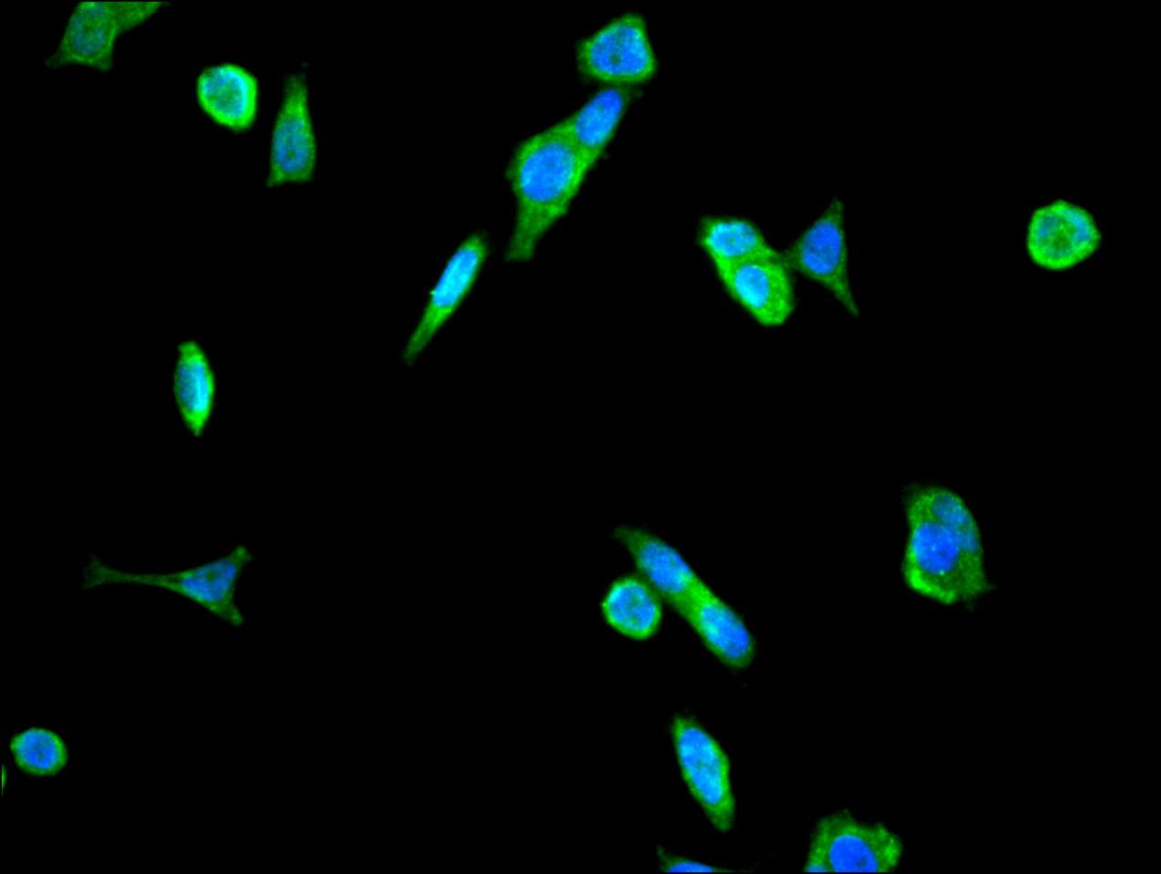
Immunofluorescence staining of MCF-7 cell with CSB-PA07889A0Rb at 1:30, counter-stained with DAPI. The cells were fixed in 4% formaldehyde and blocked in 10% normal Goat Serum. The cells were then incubated with the antibody overnight at 4C. The secondary antibody was Alexa Fluor 488-congugated AffiniPure Goat Anti-Rabbit IgG(H+L). |
|
|
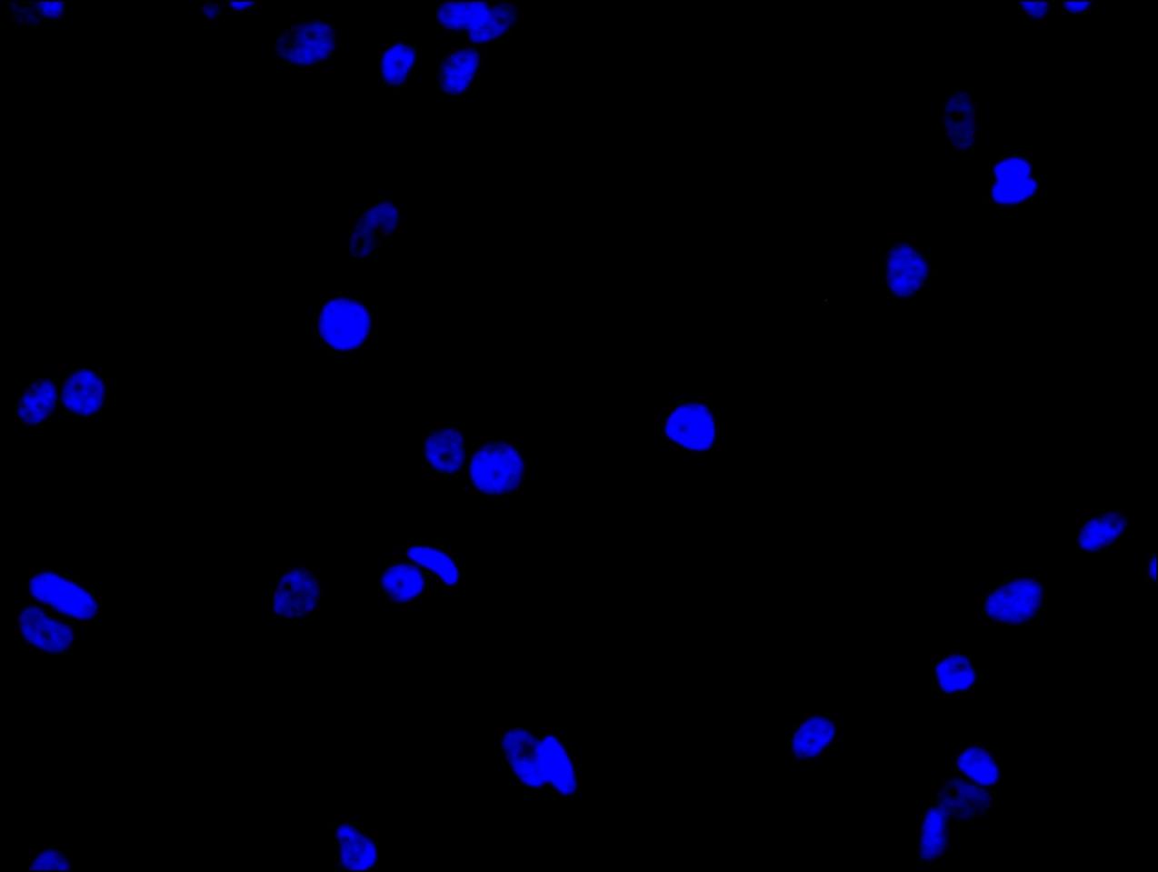
Immunofluorescence staining of MCF-7 cell with 5% goat serum, counter-stained with DAPI. The cells were fixed in 4% formaldehyde and blocked in 10% normal Goat Serum. The cells were then incubated with the antibody overnight at 4C. The secondary antibody was Alexa Fluor 488-congugated AffiniPure Goat Anti-Rabbit IgG(H+L). |
|
|
Immunofluorescence staining of U251 cell with CSB-PA07889A0Rb at 1:30, counter-stained with DAPI. The cells were fixed in 4% formaldehyde and blocked in 10% normal Goat Serum. The cells were then incubated with the antibody overnight at 4C. The secondary antibody was Alexa Fluor 488-congugated AffiniPure Goat Anti-Rabbit IgG(H+L). |
|
|
Immunofluorescence staining of U251 cell with 5% goat serum, counter-stained with DAPI. The cells were fixed in 4% formaldehyde and blocked in 10% normal Goat Serum. The cells were then incubated with the antibody overnight at 4C. The secondary antibody was Alexa Fluor 488-congugated AffiniPure Goat Anti-Rabbit IgG(H+L). |
|
|
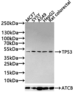
Western Blot Positive WB detected in: MCF7 whole cell lysate(20µg), SY5Y whole cell lysate(20µg), A549 whole cell lysate(20µg), HepG2 whole cell lysate(20µg), Rat colorectal tissue lysate(20µg) All lanes: TP53 antibody at 1:1000 Secondary Goat polyclonal to rabbit IgG at 1/20000 dilution Predicted band size: 44,38,39,40,34,35,30,24,25 kDa Observed band size: 53 kDa Exposure time: 120s |
Produktgarantie und fachkundiger Support
