
AKT1 Antibody, Unconjugated, Rabbit, Polyclonal
Artikelnummer:
CSB-PA15905A0RB
- Bilder (7)
| Artikelname: | AKT1 Antibody, Unconjugated, Rabbit, Polyclonal |
| Artikelnummer: | CSB-PA15905A0RB |
| Hersteller Artikelnummer: | CSB-PA15905A0Rb |
| Alternativnummer: | CSB-PA15905A0RB-100UG, CSB-PA15905A0RB-50UG |
| Hersteller: | Cusabio |
| Wirt: | Rabbit |
| Kategorie: | Antikörper |
| Applikation: | ELISA, IF, IHC, IP, WB |
| Spezies Reaktivität: | Human |
| Konjugation: | Unconjugated |
| Alternative Synonym: | AKT 1 antibody, AKT antibody, AKT1 antibody, AKT1_HUMAN antibody, C AKT antibody, cAKT antibody, MGC99656 antibody, PKB alpha antibody, PKB antibody, PKB-ALPHA antibody, PRKBA antibody, Protein Kinase B Alpha antibody, Protein kinase B antibody, Proto-oncogene c-Akt antibody, RAC Alpha antibody, RAC antibody, Rac protein kinase alpha antibody, RAC Serine/Threonine Protein Kinase antibody, RAC-alpha serine/threonine-protein kinase antibody, RAC-PK-alpha antibody, v akt murine thymoma viral oncogene homolog 1 antibody, vAKT Murine Thymoma Viral Oncogene Homolog 1 antibody |
| Klonalität: | Polyclonal |
| UniProt: | P31749 |
| Puffer: | Preservative: 0.03% Proclin 300<br />Constituents: 50% Glycerol, 0.01M PBS, PH 7.4 |
| Reinheit: | >95%, Protein G purified |
| Formulierung: | Liquid |
| Target-Kategorie: | AKT1 |
| Application Verdünnung: | Recommended dilution: WB:1:500-1:5000, IHC:1:100-1:1000, IF:1:200-1:500, IP:1:200-1:2000 |
|
|
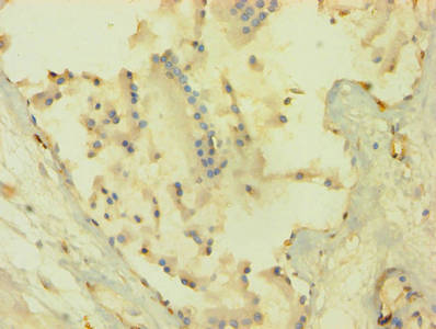
Immunohistochemistry of paraffin-embedded human prostate tissue using CSB-PA15905A0Rb at dilution of 1:100 |
|
|
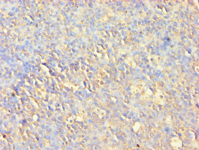
Immunohistochemistry of paraffin-embedded human tonsil tissue using CSB-PA15905A0Rb at dilution of 1:100 |
|
|
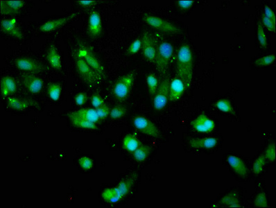
Immunofluorescence staining of Hela cells with CSB-PA15905A0Rb ,at 1:200, counter-stained with DAPI. The cells were fixed in 4% formaldehyde, permeabilized using 0.2% Triton X-100 and blocked in 10% normal Goat Serum. The cells were then incubated with the antibody overnight at 4°,C. The secondary antibody was Alexa Fluor 488-congugated AffiniPure Goat Anti-Rabbit IgG(H+L). |
|
|
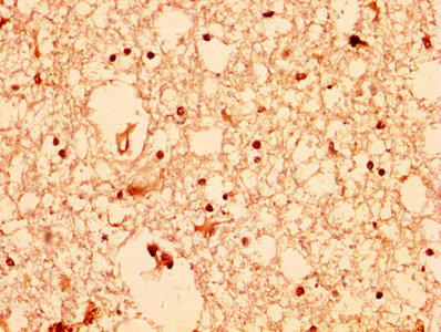
IHC image of CSB-PA15905A0Rb diluted at 1:200 and staining in paraffin-embedded human brain tissue performed on a Leica BondTM system. After dewaxing and hydration, antigen retrieval was mediated by high pressure in a citrate buffer (pH 6.0). Section was blocked with 10% normal goat serum 30min at RT. Then primary antibody (1% BSA) was incubated at 4°,C overnight. The primary is detected by a biotinylated secondary antibody and visualized using an HRP conjugated SP system. |
|
|
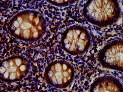
IHC image of CSB-PA15905A0Rb ,diluted at 1:700 and staining in paraffin-embedded human appendix tissue performed on a Leica BondTM system. After dewaxing and hydration, antigen retrieval was mediated by high pressure in a citrate buffer (pH 6.0). Section was blocked with 10% normal goat serum 30min at RT. Then primary antibody (1% BSA) was incubated at 4°,C overnight. The primary is detected by a biotinylated secondary antibody and visualized using an HRP conjugated SP system. |
|
|
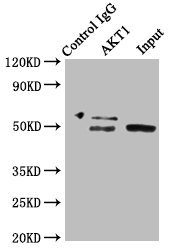
Immunoprecipitating AKT1 in HepG2 whole cell lysate Lane 1: Rabbit control IgG instead of CSB-PA15905A0Rb in HepG2 whole cell lysate. For western blotting, a HRP-conjugated Protein G antibody was used as the secondary antibody (1/2000) Lane 2: CSB-PA15905A0Rb (8µg) + HepG2 whole cell lysate (500µg) Lane 3: HepG2 whole cell lysate (20µg) |
|
|
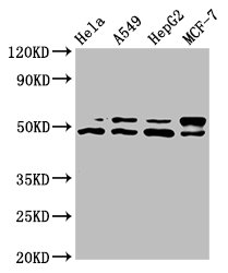
Western Blot Positive WB detected in: Hela whole cell lysate, A549 whole cell lysate, HepG2 whole cell lysate, MCF-7 whole cell lysate All lanes: AKT1 antibody at 7.4µg/ml Secondary Goat polyclonal to rabbit IgG at 1/50000 dilution Predicted band size: 56, 49 kDa Observed band size: 56, 49 kDa |
Produktgarantie und fachkundiger Support
