
SOX2 Antibody, Unconjugated, Rabbit, Polyclonal
Artikelnummer:
CSB-PA16539A0RB
- Bilder (7)


| Artikelname: | SOX2 Antibody, Unconjugated, Rabbit, Polyclonal |
| Artikelnummer: | CSB-PA16539A0RB |
| Hersteller Artikelnummer: | CSB-PA16539A0Rb |
| Alternativnummer: | CSB-PA16539A0RB-100UG, CSB-PA16539A0RB-50UG |
| Hersteller: | Cusabio |
| Wirt: | Rabbit |
| Kategorie: | Antikörper |
| Applikation: | ELISA, IF, IHC, WB |
| Spezies Reaktivität: | Human, Mouse |
| Konjugation: | Unconjugated |
| Alternative Synonym: | ANOP3 antibody, cb236 antibody, Delta EF2a antibody, lcc antibody, MCOPS3 antibody, MGC148683 antibody, MGC2413 antibody, RGD1565646 antibody, Sex determining region Y box 2 antibody, SOX 2 antibody, Sox2 antibody, SOX2_HUMAN antibody, SRY (sex determining region Y) box 2 antibody, SRY box containing gene 2 antibody, SRY related HMG box 2 antibody, SRY related HMG box gene 2 antibody, SRY-box 2 antibody, Transcription factor SOX 2 antibody, Transcription factor SOX-2 antibody, ysb antibody |
| Klonalität: | Polyclonal |
| UniProt: | P48431 |
| Puffer: | Preservative: 0.03% Proclin 300<br />Constituents: 50% Glycerol, 0.01M PBS, PH 7.4 |
| Reinheit: | >95%, Protein G purified |
| Formulierung: | Liquid |
| Target-Kategorie: | SOX2 |
| Application Verdünnung: | Recommended dilution: WB:1:500-1:5000, IHC:1:20-1:200, IF:1:50-1:200 |
|
|
IHC image of CSB-PA16539A0Rb diluted at 1:500 and staining in paraffin-embedded human lung cancer performed on a Leica BondTM system. After dewaxing and hydration, antigen retrieval was mediated by high pressure in a citrate buffer (pH 6.0). Section was blocked with 10% normal goat serum 30min at RT. Then primary antibody (1% BSA) was incubated at 4C overnight. The primary is detected by a biotinylated secondary antibody and visualized using an HRP conjugated SP system. |
|
|
Immunofluorescence staining of HepG2 cells with CSB-PA16539A0Rb at 1:166, counter-stained with DAPI. The cells were fixed in 4% formaldehyde, permeabilized using 0.2% Triton X-100 and blocked in 10% normal Goat Serum. The cells were then incubated with the antibody overnight at 4C. The secondary antibody was Alexa Fluor 488-congugated AffiniPure Goat Anti-Rabbit IgG(H+L). |
|
|
IHC image of CSB-PA16539A0Rb diluted at 1:500 and staining in paraffin-embedded human breast cancer performed on a Leica BondTM system. After dewaxing and hydration, antigen retrieval was mediated by high pressure in a citrate buffer (pH 6.0). Section was blocked with 10% normal goat serum 30min at RT. Then primary antibody (1% BSA) was incubated at 4C overnight. The primary is detected by a biotinylated secondary antibody and visualized using an HRP conjugated SP system. |
|
|
Western blot All lanes: Transcription factor SOX-2 antibody at 2ug/ml + Mouse kidney tissue Secondary Goat polyclonal to rabbit IgG at 1/15000 dilution Predicted band size: 34 kDa Observed band size: 34 kDa |
|
|
Western Blot All lanes: Transcription factor SOX-2 antibody at 6ug/ml + 293T whole cell lysate Secondary Goat polyclonal to rabbit IgG at 1/15000 dilution Predicted band size: 34 kDa Observed band size: 34 kDa |
|
|
Western Blot Positive WB detected in: Mouse brain tissue All lanes: SOX2 antibody at 3.5ug/ml Secondary Goat polyclonal to rabbit IgG at 1/50000 dilution Predicted band size: 34 kDa Observed band size: 34 kDa |
|
|
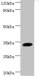
Western Blot Positive WB detected in: Zebrafish 40ug, 20ug, 10ug All lanes: Sox2 antibody at 3ug/ml Secondary Goat polyclonal to rabbit IgG at 1/50000 dilution Predicted band size: 35 kDa Observed band size: 35 kDa |
Produktgarantie und fachkundiger Support
