
MRGPRX2 Antibody, Unconjugated, Rabbit, Polyclonal
Artikelnummer:
CSB-PA839361LA01HU
- Bilder (9)


| Artikelname: | MRGPRX2 Antibody, Unconjugated, Rabbit, Polyclonal |
| Artikelnummer: | CSB-PA839361LA01HU |
| Hersteller Artikelnummer: | CSB-PA839361LA01HU |
| Alternativnummer: | CSB-PA839361LA01HU-100UG, CSB-PA839361LA01HU-50UG |
| Hersteller: | Cusabio |
| Wirt: | Rabbit |
| Kategorie: | Antikörper |
| Applikation: | ELISA, IF, WB |
| Spezies Reaktivität: | Human, Mouse |
| Konjugation: | Unconjugated |
| Alternative Synonym: | G protein coupled receptor MRGX 2 antibody, G protein coupled receptor MRGX2 antibody, Mas related G protein coupled receptor member X2 antibody, Mas related gene MRGX 2 antibody, Mas related gene MRGX2 antibody, MAS related GPR member X2 antibody, Mas-related G-protein coupled receptor member X antibody, Mas-related G-protein coupled receptor member X2 antibody, MRGPRX 2 antibody, MRGPRX2 antibody, MRGX 2 antibody, MRGX2 antibody, MRGX2_HUMAN antibody |
| Klonalität: | Polyclonal |
| UniProt: | Q96LB1 |
| Puffer: | Preservative: 0.03% Proclin 300<br />Constituents: 50% Glycerol, 0.01M PBS, PH 7.4 |
| Reinheit: | >95%, Protein G purified |
| Formulierung: | Liquid |
| Target-Kategorie: | MRGPRX2 |
| Application Verdünnung: | Recommended dilution: WB:1:1000-1:5000, IF:1:50-1:200 |
|
|
|
|
|
|
|
|
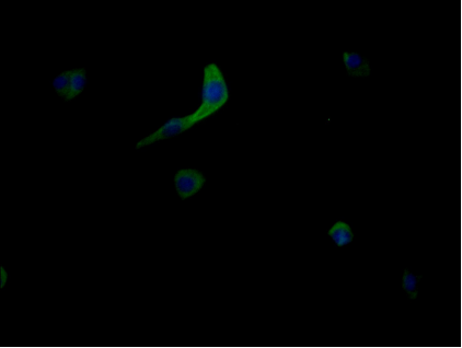
Immunofluorescent analysis of MCF-7 cells using CSB-PA839361LA01HU at dilution of 1:100 and Alexa Fluor 488-congugated AffiniPure Goat Anti-Rabbit IgG(H+L) |
|
|
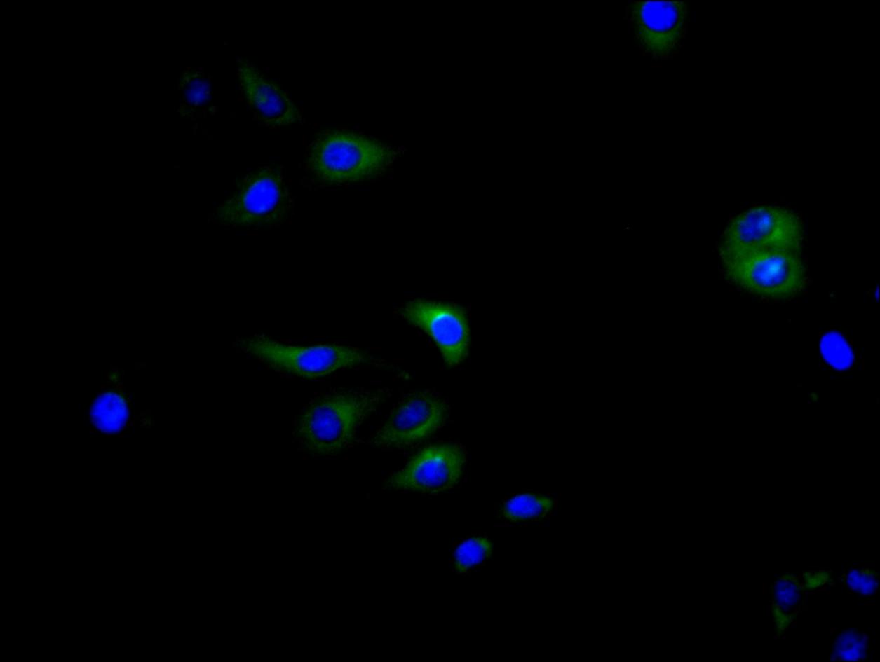
Immunofluorescence staining of Hela cell with CSB-PA839361LA01HU at 1:60, counter-stained with DAPI. The cells were fixed in 4% formaldehyde and blocked in 10% normal Goat Serum. The cells were then incubated with the antibody overnight at 4C. The secondary antibody was Alexa Fluor 488-congugated AffiniPure Goat Anti-Rabbit IgG(H+L). |
|
|
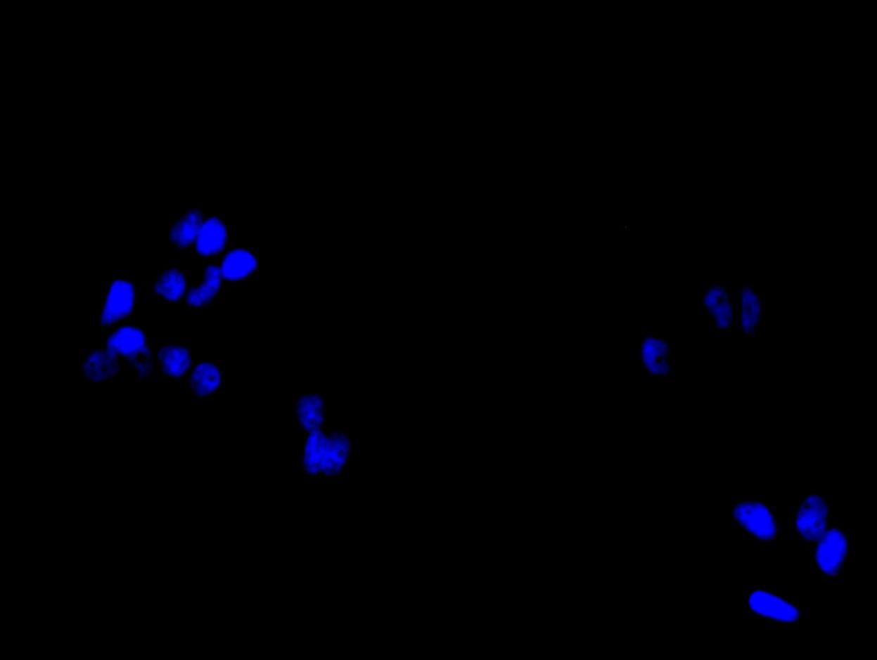
Immunofluorescence staining of Hela cell with 5% goat serum, counter-stained with DAPI. The cells were fixed in 4% formaldehyde and blocked in 10% normal Goat Serum. The cells were then incubated with the antibody overnight at 4C. The secondary antibody was Alexa Fluor 488-congugated AffiniPure Goat Anti-Rabbit IgG(H+L). |
|
|
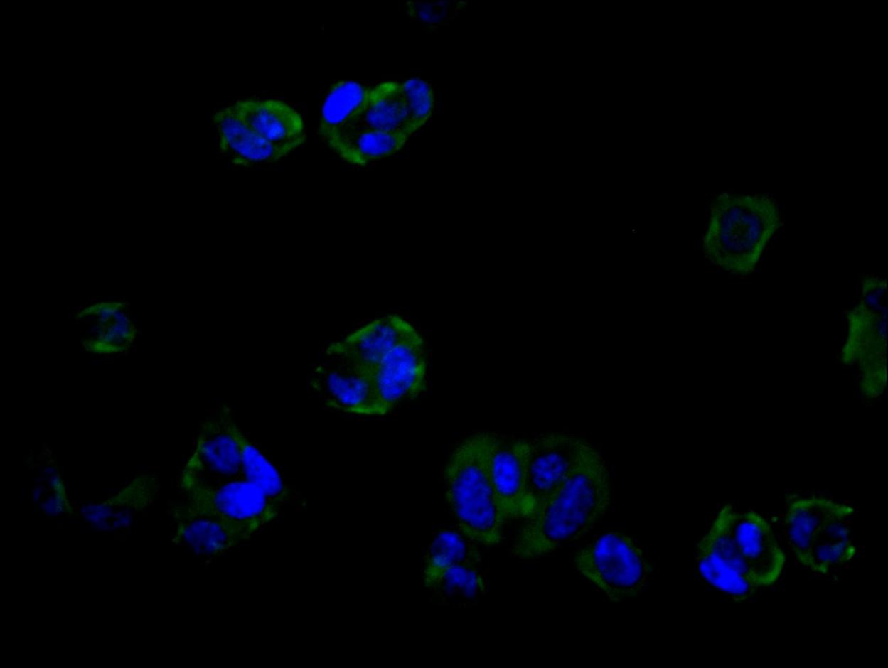
Immunofluorescence staining of MCF-7 cell with CSB-PA839361LA01HU at 1:60, counter-stained with DAPI. The cells were fixed in 4% formaldehyde and blocked in 10% normal Goat Serum. The cells were then incubated with the antibody overnight at 4C. The secondary antibody was Alexa Fluor 488-congugated AffiniPure Goat Anti-Rabbit IgG(H+L). |
|
|
Immunofluorescence staining of MCF-7 cell with 5% goat serum, counter-stained with DAPI. The cells were fixed in 4% formaldehyde and blocked in 10% normal Goat Serum. The cells were then incubated with the antibody overnight at 4C. The secondary antibody was Alexa Fluor 488-congugated AffiniPure Goat Anti-Rabbit IgG(H+L). |
|
|
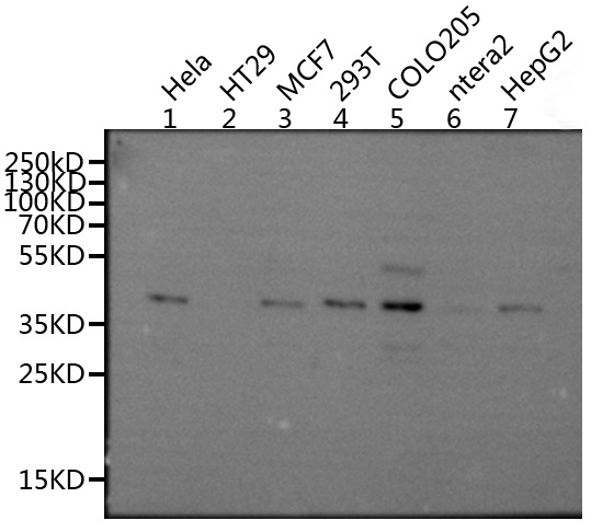
Western Blot Positive WB detected in: Hela whole cell lysate,MCF7 whole cell lysate, 293T whole cell lysate, COLO205 whole cell lysate, HepG2 whole cell lysate All lanes: MRGPRX2 antibody at 1:1000 Secondary Goat polyclonal to rabbit IgG at 1/50000 dilution Predicted band size: 38 kDa Observed band size: 38 kDa |
|
|
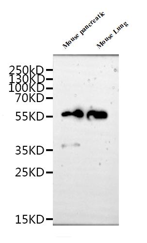
Western Blot Positive WB detected in: Mouse pancreatic tissue lysate, Mouse Lung tissue lysate All lanes: MRGPRX2 antibody at 1:1000 Secondary Goat polyclonal to rabbit IgG at 1/50000 dilution Predicted band size: 40 kDa Observed band size: 40, 55 kDa |
Produktgarantie und fachkundiger Support
