
CLCC1 Antibody, Unconjugated, Rabbit, Polyclonal
Artikelnummer:
CSB-PA857024LA01HU
- Bilder (5)
| Artikelname: | CLCC1 Antibody, Unconjugated, Rabbit, Polyclonal |
| Artikelnummer: | CSB-PA857024LA01HU |
| Hersteller Artikelnummer: | CSB-PA857024LA01HU |
| Alternativnummer: | CSB-PA857024LA01HU-100UG, CSB-PA857024LA01HU-50UG |
| Hersteller: | Cusabio |
| Wirt: | Rabbit |
| Kategorie: | Antikörper |
| Applikation: | ELISA, IF, IHC, IP, WB |
| Spezies Reaktivität: | Human |
| Konjugation: | Unconjugated |
| Alternative Synonym: | CLCC1 antibody, KIAA0761 antibody, MCLCChloride channel CLIC-like protein 1 antibody, Mid-1-related chloride channel protein 1 antibody |
| Klonalität: | Polyclonal |
| UniProt: | Q96S66 |
| Puffer: | Preservative: 0.03% Proclin 300<br />Constituents: 50% Glycerol, 0.01M PBS, pH 7.4 |
| Reinheit: | >95%, Protein G purified |
| Formulierung: | Liquid |
| Target-Kategorie: | CLCC1 |
| Application Verdünnung: | Recommended dilution: WB:1:500-1:5000, IHC:1:200-1:500, IF:1:50-1:200, IP:1:200-1:2000 |
|
|
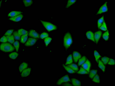
Immunofluorescence staining of Hela cells with CSB-PA857024LA01HU at 1:133, counter-stained with DAPI. The cells were fixed in 4% formaldehyde, permeabilized using 0.2% Triton X-100 and blocked in 10% normal Goat Serum. The cells were then incubated with the antibody overnight at 4°,C. The secondary antibody was Alexa Fluor 488-congugated AffiniPure Goat Anti-Rabbit IgG(H+L). |
|
|
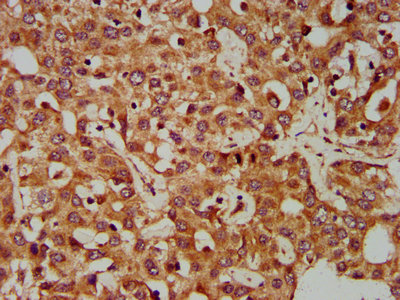
IHC image of CSB-PA857024LA01HU diluted at 1:400 and staining in paraffin-embedded human liver cancer performed on a Leica BondTM system. After dewaxing and hydration, antigen retrieval was mediated by high pressure in a citrate buffer (pH 6.0). Section was blocked with 10% normal goat serum 30min at RT. Then primary antibody (1% BSA) was incubated at 4°,C overnight. The primary is detected by a biotinylated secondary antibody and visualized using an HRP conjugated SP system. |
|
|
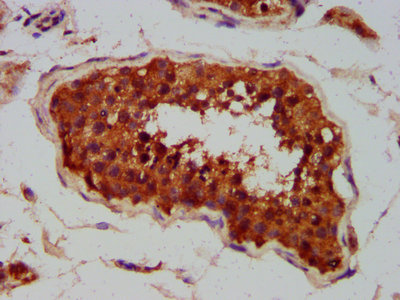
IHC image of CSB-PA857024LA01HU diluted at 1:400 and staining in paraffin-embedded human testis tissue performed on a Leica BondTM system. After dewaxing and hydration, antigen retrieval was mediated by high pressure in a citrate buffer (pH 6.0). Section was blocked with 10% normal goat serum 30min at RT. Then primary antibody (1% BSA) was incubated at 4°,C overnight. The primary is detected by a biotinylated secondary antibody and visualized using an HRP conjugated SP system. |
|
|
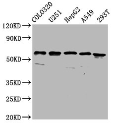
Western Blot Positive WB detected in: Colo320 whole cell lysate, U251 whole cell lysate, HepG2 whole cell lysate, A549 whole cell lysate, 293T whole cell lysate All lanes: CLCC1 antibody at 3.7µg/ml Secondary Goat polyclonal to rabbit IgG at 1/50000 dilution Predicted band size: 63, 57, 48, 40 kDa Observed band size: 63 kDa |
|
|
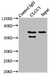
Immunoprecipitating CLCC1 in HepG2 whole cell lysate Lane 1: Rabbit control IgG instead of CSB-PA857024LA01HU in HepG2 whole cell lysate. For western blotting, a HRP-conjugated Protein G antibody was used as the secondary antibody (1/2000) Lane 2: CSB-PA857024LA01HU (6µg) + HepG2 whole cell lysate (500µg) Lane 3: HepG2 whole cell lysate (20µg) |
Produktgarantie und fachkundiger Support
