
WWOX Antibody, Unconjugated, Rabbit, Polyclonal
Artikelnummer:
CSB-PA873704LA01HU
- Bilder (6)
| Artikelname: | WWOX Antibody, Unconjugated, Rabbit, Polyclonal |
| Artikelnummer: | CSB-PA873704LA01HU |
| Hersteller Artikelnummer: | CSB-PA873704LA01HU |
| Alternativnummer: | CSB-PA873704LA01HU-100UG, CSB-PA873704LA01HU-50UG |
| Hersteller: | Cusabio |
| Wirt: | Rabbit |
| Kategorie: | Antikörper |
| Applikation: | ELISA, IF, IHC, IP, WB |
| Spezies Reaktivität: | Human, Rat |
| Konjugation: | Unconjugated |
| Alternative Synonym: | 5330426P09Rik antibody, 9030416C10Rik antibody, Aberrant WW domain-containing oxidoreductase antibody, D16S432E antibody, EC 1.1.1.- antibody, EIEE28 antibody, FOR antibody, FRA16D antibody, Fragile site FRA16D oxidoreductase antibody, Fragile site FRA16D Oxireductase antibody, HHCMA56 antibody, MGC55975 antibody, PRO0128 antibody, Putative oxidoreductase antibody, SCAR12 antibody, SDR41C1 antibody, Short chain dehydrogenase/reductase family 41C, member 1 antibody, WOX1 antibody, WW domain containing oxidoreductase antibody, WW domain-containing oxidoreductase antibody, WW domain-containing protein WWOX antibody, wwox antibody, WWOX_HUMAN antibody, zgc:55975 antibody |
| Klonalität: | Polyclonal |
| UniProt: | Q9NZC7 |
| Puffer: | Preservative: 0.03% Proclin 300<br />Constituents: 50% Glycerol, 0.01M PBS, pH 7.4 |
| Reinheit: | >95%, Protein G purified |
| Formulierung: | Liquid |
| Target-Kategorie: | WWOX |
| Application Verdünnung: | Recommended dilution: WB:1:500-1:5000, IHC:1:200-1:500, IF:1:50-1:200, IP:1:200-1:2000 |
|
|
Immunofluorescence staining of A549 cells with CSB-PA873704LA01HU at 1:100, counter-stained with DAPI. The cells were fixed in 4% formaldehyde, permeabilized using 0.2% Triton X-100 and blocked in 10% normal Goat Serum. The cells were then incubated with the antibody overnight at 4°,C. The secondary antibody was Alexa Fluor 488-congugated AffiniPure Goat Anti-Rabbit IgG(H+L). |
|
|
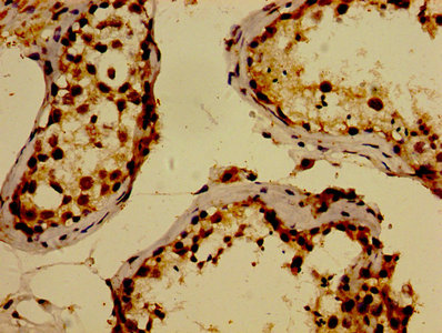
IHC image of CSB-PA873704LA01HU diluted at 1:300 and staining in paraffin-embedded human testis tissue performed on a Leica BondTM system. After dewaxing and hydration, antigen retrieval was mediated by high pressure in a citrate buffer (pH 6.0). Section was blocked with 10% normal goat serum 30min at RT. Then primary antibody (1% BSA) was incubated at 4°,C overnight. The primary is detected by a biotinylated secondary antibody and visualized using an HRP conjugated SP system. |
|
|
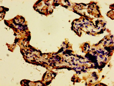
IHC image of CSB-PA873704LA01HU diluted at 1:300 and staining in paraffin-embedded human placenta tissue performed on a Leica BondTM system. After dewaxing and hydration, antigen retrieval was mediated by high pressure in a citrate buffer (pH 6.0). Section was blocked with 10% normal goat serum 30min at RT. Then primary antibody (1% BSA) was incubated at 4°,C overnight. The primary is detected by a biotinylated secondary antibody and visualized using an HRP conjugated SP system. |
|
|
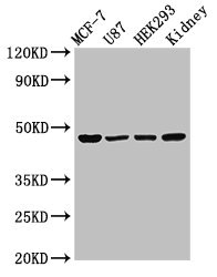
Western Blot Positive WB detected in: MCF-7 whole cell lysate, U87 whole cell lysate, HEK293 whole cell lysate, Rat kidney tissue All lanes: WWOX antibody at 3µg/ml Secondary Goat polyclonal to rabbit IgG at 1/50000 dilution Predicted band size: 47, 42, 22, 5, 27, 36, 24 kDa Observed band size: 47 kDa |
|
|
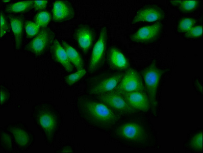
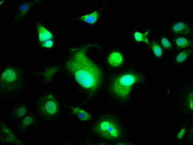
Immunofluorescence staining of MCF-7 cells with CSB-PA873704LA01HU at 1:100, counter-stained with DAPI. The cells were fixed in 4% formaldehyde, permeabilized using 0.2% Triton X-100 and blocked in 10% normal Goat Serum. The cells were then incubated with the antibody overnight at 4°,C. The secondary antibody was Alexa Fluor 488-congugated AffiniPure Goat Anti-Rabbit IgG(H+L). |
|
|
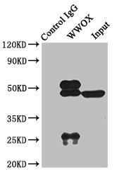
Immunoprecipitating WWOX in Rat kidney tissue Lane 1: Rabbit control IgG instead of CSB-PA873704LA01HU in Rat kidney tissue. For western blotting, a HRP-conjugated light chain specific antibody was used as the secondary antibody (1/50000) Lane 2: CSB-PA873704LA01HU (8µg) + Rat kidney tissue (500µg) Lane 3: Rat kidney tissue (10µg) |
Produktgarantie und fachkundiger Support
