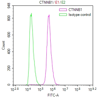
Overlay Peak curve showing Hela cells stained with CSB-RA006169MA1HU (red line) at 1:100. The cells were fixed in 4% formaldehyde (15min) and permeated by 0.2% TritonX-100 for 10min. Then 10% normal goat serum to block non-specific protein-protein interactions followed by the antibody (1ug/1*106cells) for 45min at 4°C. The secondary antibody used was FITC-conjugated Goat Anti-Rabbit IgG(H+L) at 1/200 dilution for 35 min at 4C. Isotype control antibody (green line) was rabbit IgG1 (1µg/1*106cells) used under the same conditions. Acquisition of >10,000 events was performed.
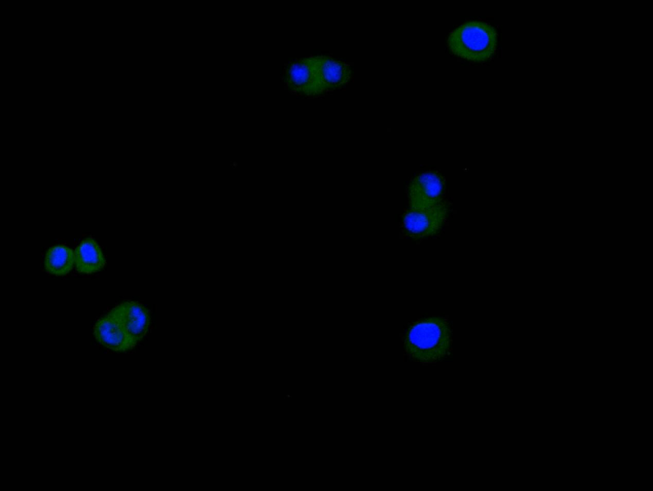
Immunofluorescence staining of Hela cell with CSB-RA006169MA1HU at 1:200, counter-stained with DAPI. The cells were fixed in 4% formaldehyde and blocked in 10% normal Goat Serum. The cells were then incubated with the antibody overnight at 4C. The secondary antibody was Alexa Fluor 488-congugated AffiniPure Goat Anti-rabbit IgG(H+L) .
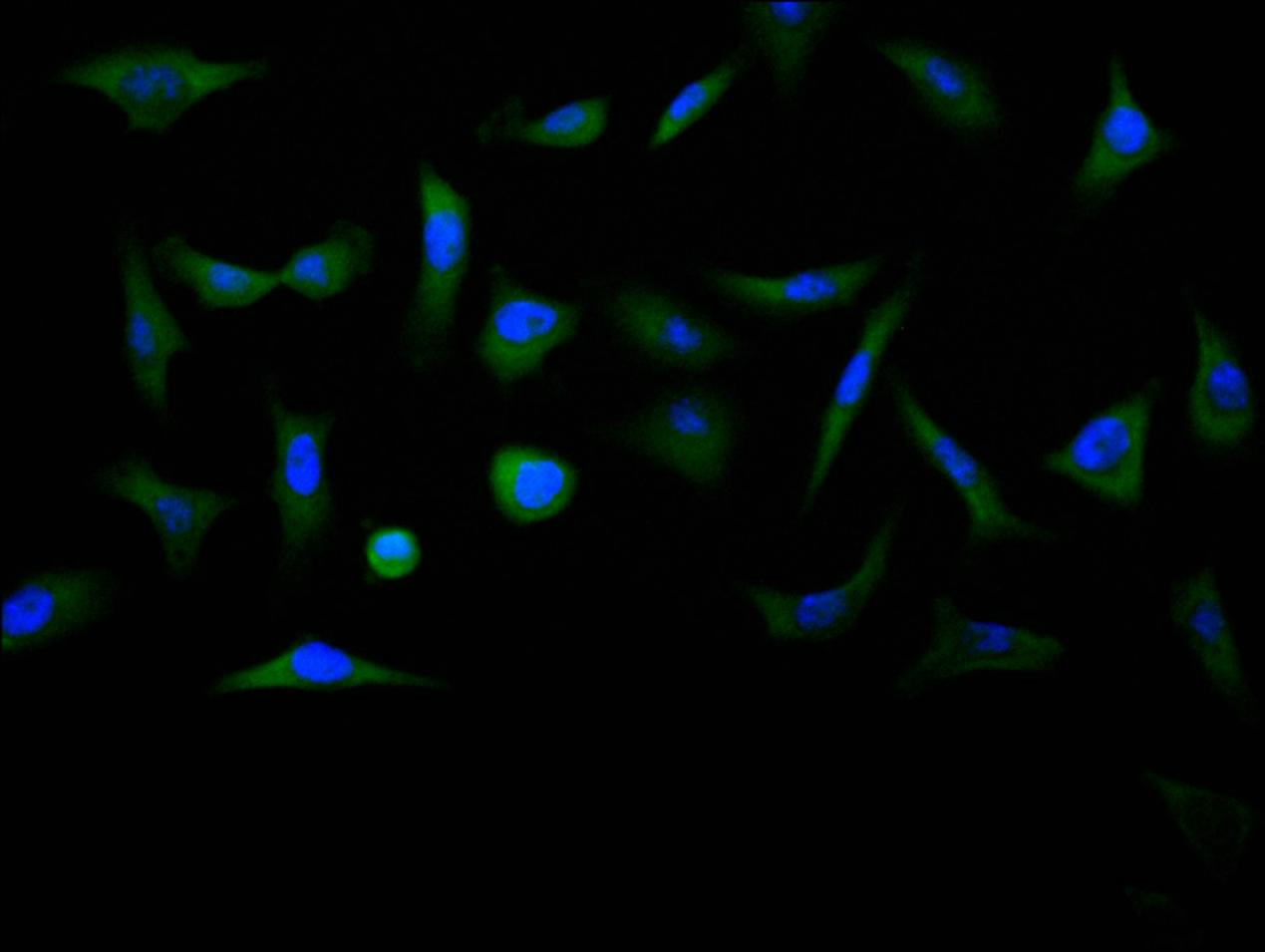
Immunofluorescence staining of MCF7 cell with CSB-RA006169MA1HU at 1:200, counter-stained with DAPI. The cells were fixed in 4% formaldehyde and blocked in 10% normal Goat Serum. The cells were then incubated with the antibody overnight at 4C. The secondary antibody was Alexa Fluor 488-congugated AffiniPure Goat Anti-rabbit IgG(H+L) .
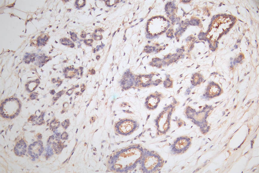
IHC image of CSB-RA006169MA1HU diluted at 1:100 and staining in paraffin-embedded human breast tissue performed on a Leica BondTM system. After dewaxing and hydration, antigen retrieval was mediated by high pressure in a citrate buffer (pH 6.0) . Section was blocked with 10% normal goat serum 30min at RT. Then primary antibody (1% BSA) was incubated at 4C overnight. The primary is detected by a Goat anti-rabbit polymer IgG labeled by HRP and visualized using 0.05% DAB.
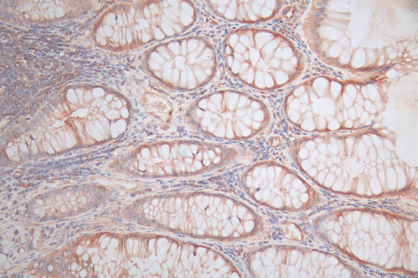
IHC image of CSB-RA006169MA1HU diluted at 1:100 and staining in paraffin-embedded human rectal cancer performed on a Leica BondTM system. After dewaxing and hydration, antigen retrieval was mediated by high pressure in a citrate buffer (pH 6.0) . Section was blocked with 10% normal goat serum 30min at RT. Then primary antibody (1% BSA) was incubated at 4C overnight. The primary is detected by a Goat anti-rabbit polymer IgG labeled by HRP and visualized using 0.05% DAB.
* Mehrwertsteuer und Versandkosten nicht enthalten. Irrtümer und Preisänderungen vorbehalten