
HSPB1 Recombinant Monoclonal Antibody, Clone: [10C11], Unconjugated, Rabbit
Artikelnummer:
CSB-RA010833A0HU
- Bilder (6)
| Artikelname: | HSPB1 Recombinant Monoclonal Antibody, Clone: [10C11], Unconjugated, Rabbit |
| Artikelnummer: | CSB-RA010833A0HU |
| Hersteller Artikelnummer: | CSB-RA010833A0HU |
| Alternativnummer: | CSB-RA010833A0HU-100UL, CSB-RA010833A0HU-50UL |
| Hersteller: | Cusabio |
| Wirt: | Rabbit |
| Kategorie: | Antikörper |
| Applikation: | ELISA, FC, IF, IHC, IP, WB |
| Spezies Reaktivität: | Human |
| Konjugation: | Unconjugated |
| Alternative Synonym: | Heat shock protein beta-1, HspB1, 28 kDa heat shock protein, Estrogen-regulated 24 kDa protein, Heat shock 27 kDa protein, HSP 27, Stress-responsive protein 27, SRP27, HSPB1, HSP27, HSP28 |
| Klonalität: | Monoclonal |
| Klon-Bezeichnung: | [10C11] |
| UniProt: | P04792 |
| Puffer: | Rabbit IgG in 10mM phosphate buffered saline , pH 7.4, 150mM sodium chloride, 0.05% BSA, 0.02% sodium azide and 50% glycerol. |
| Reinheit: | Affinity-chromatography |
| Formulierung: | Liquid |
| Target-Kategorie: | HSPB1 |
| Antibody Type: | Recombinant Antibody |
| Application Verdünnung: | Recommended dilution: WB:1:500-1:5000, IHC:1:50-1:200, IF:1:20-1:200, IP:1:200-1:1000 |
|
|
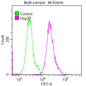
Overlay histogram showing Hela cells stained with CSB-RA010833A0HU (red line) at 1:50. The cells were fixed with 70% Ethylalcohol (18h) and then permeabilized with 0.3% Triton X-100 for 2 min. The cells were then incubated in 1x PBS /10% normal goat serum to block non-specific protein-protein interactions followed by primary antibody for 1 h at 4°C. The secondary antibody used was FITC goat anti-rabbit IgG (H+L) at 1/200 dilution for 1 h at 4°C. Control antibody (green line) was used under the same conditions. Acquisition of >10,000 events was performed. |
|
|
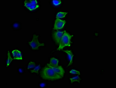
Immunofluorescence staining of MCF-7 cells with CSB-RA010833A0HU at 1:20, counter-stained with DAPI. The cells were fixed in 4% formaldehyde, permeabilized using 0.2% Triton X-100 and blocked in 10% normal Goat Serum. The cells were then incubated with the antibody overnight at 4°C. The secondary antibody was Alexa Fluor 488-congugated AffiniPure Goat Anti-Rabbit IgG (H+L) . |
|
|
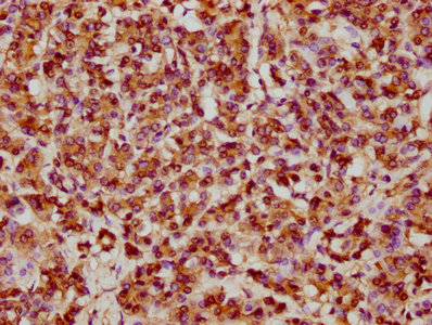
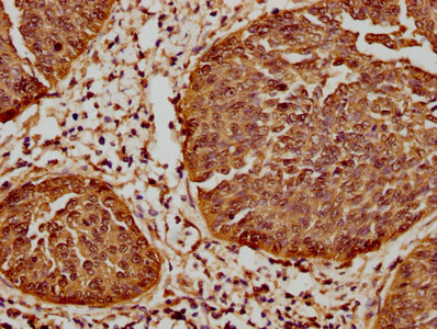
IHC image of CSB-RA010833A0HU diluted at 1:61.9 and staining in paraffin-embedded human pancreatic tissue performed on a Leica BondTM system. After dewaxing and hydration, antigen retrieval was mediated by high pressure in a citrate buffer (pH 6.0) . Section was blocked with 10% normal goat serum 30min at RT. Then primary antibody (1% BSA) was incubated at 4°C overnight. The primary is detected by a biotinylated secondary antibody and visualized using an HRP conjugated SP system. |
|
|
IHC image of CSB-RA010833A0HU diluted at 1:61.9 and staining in paraffin-embedded human cervical cancer performed on a Leica BondTM system. After dewaxing and hydration, antigen retrieval was mediated by high pressure in a citrate buffer (pH 6.0) . Section was blocked with 10% normal goat serum 30min at RT. Then primary antibody (1% BSA) was incubated at 4°C overnight. The primary is detected by a biotinylated secondary antibody and visualized using an HRP conjugated SP system. |
|
|
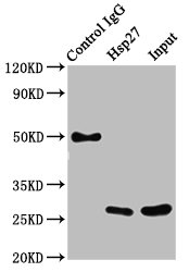
Immunoprecipitating Hsp27 in Hela whole cell lysate Lane 1: Rabbit control IgG instead of CSB-RA010833A0HU in Hela whole cell lysate.For western blotting, a HRP-conjugated Protein G antibody was used as the secondary antibody (1/2000) Lane 2: CSB-RA010833A0HU (3µg) + Hela whole cell lysate (500µg) Lane 3: Hela whole cell lysate (20µg) |
|
|
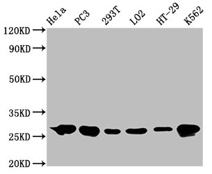
Western Blot Positive WB detected in: Hela whole cell lysate, PC3 whole cell lysate, 293T whole cell lysate, LO2 whole cell lysate, HT-29 whole cell lysate, K562 whole cell lysate All lanes: Hsp27 antibody at 0.62µg/ml Secondary Goat polyclonal to rabbit IgG at 1/50000 dilution Predicted band size: 23 KDa Observed band size: 27 KDa |
Produktgarantie und fachkundiger Support
