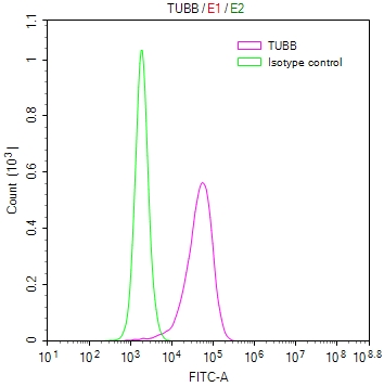
Overlay Peak curve showing K562 cells stained with CSB-RA025318MA1HU (red line) at 1:100.The cells were fixed in 4% formaldehyde and permeated by 0.2% TritonX-100. Then 10% normal goat serum to block non-specific protein-protein interactions followed by the antibody (1ug/1*106cells) for 45min at 4°C. The secondary antibody used was FITC-conjugated Goat Anti-human IgG(H+L) at 1:200 dilution for 35min at 4°C.Control antibody (green line) was human IgG (1ug/1*106cells) used under the same conditions. Acquisition of >10,000 events was performed.
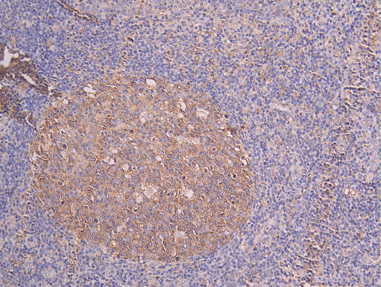
IHC image of CSB-RA008615MA1HU diluted at 1:200 and staining in paraffin-embedded human tonsil tissue performed on a Leica BondTM system. After dewaxing and hydration, antigen retrieval was mediated by high pressure in a citrate buffer (pH 6.0) . Section was blocked with 10% normal goat serum 30min at RT. Then primary antibody (1% BSA) was incubated at 4C overnight. The primary is detected by a Goat anti-human polymer IgG labeled by HRP and visualized using 0.05% DAB.
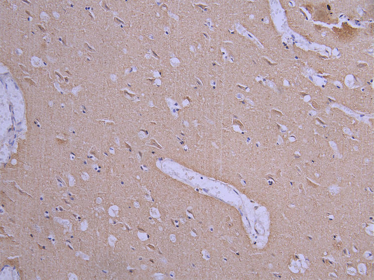
IHC image of CSB-RA008615MA1HU diluted at 1:200 and staining in paraffin-embedded human brain tissue performed on a Leica BondTM system. After dewaxing and hydration, antigen retrieval was mediated by high pressure in a citrate buffer (pH 6.0) . Section was blocked with 10% normal goat serum 30min at RT. Then primary antibody (1% BSA) was incubated at 4C overnight. The primary is detected by a Goat anti-human polymer IgG labeled by HRP and visualized using 0.05% DAB.
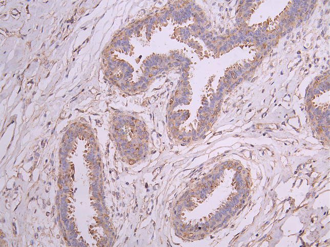
IHC image of CSB-RA008615MA1HU diluted at 1:200 and staining in paraffin-embedded human breast cancer performed on a Leica BondTM system. After dewaxing and hydration, antigen retrieval was mediated by high pressure in a citrate buffer (pH 6.0) . Section was blocked with 10% normal goat serum 30min at RT. Then primary antibody (1% BSA) was incubated at 4C overnight. The primary is detected by a Goat anti-human polymer IgG labeled by HRP and visualized using 0.05% DAB.
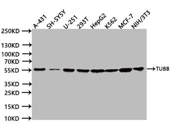
Western BlotPositive WB detected in:A431 whole cell lysate (20µg) , SH-SY5Y whole cell lysate (20µg) , U251 whole cell lysate (20µg) , 293T whole cell lysate (20µg) , HepG2 whole cell lysate (20µg) , K562 whole cell lysate (20µg) , MCF7 whole cell lysate (20µg) , NIH/3T3 whole cell lysate (20µg) All lanes: TUBB antibody at 1:1000SecondaryGoat polyclonal to human IgG at 1/40000 dilutionPredicted band size:49 kDaObserved band size:55 kDaExposure time:5s
* Mehrwertsteuer und Versandkosten nicht enthalten. Irrtümer und Preisänderungen vorbehalten