
CDK6 Recombinant Monoclonal Antibody, Clone: [8G3], Unconjugated, Rabbit
Artikelnummer:
CSB-RA555745A0HU
- Bilder (8)
| Artikelname: | CDK6 Recombinant Monoclonal Antibody, Clone: [8G3], Unconjugated, Rabbit |
| Artikelnummer: | CSB-RA555745A0HU |
| Hersteller Artikelnummer: | CSB-RA555745A0HU |
| Alternativnummer: | CSB-RA555745A0HU-100UL, CSB-RA555745A0HU-50UL |
| Hersteller: | Cusabio |
| Wirt: | Rabbit |
| Kategorie: | Antikörper |
| Applikation: | ELISA, IF, IHC, IP, WB |
| Spezies Reaktivität: | Human |
| Konjugation: | Unconjugated |
| Alternative Synonym: | Cyclin-dependent kinase 6 (EC 2.7.11.22) (Cell division protein kinase 6) (Serine/threonine-protein kinase PLSTIRE) , CDK6, CDKN6 |
| Klonalität: | Monoclonal |
| Klon-Bezeichnung: | [8G3] |
| UniProt: | Q00534 |
| Puffer: | Rabbit IgG in 10mM phosphate buffered saline , pH 7.4, 150mM sodium chloride, 0.05% BSA, 0.02% sodium azide and 50% glycerol. |
| Reinheit: | Affinity-chromatography |
| Formulierung: | Liquid |
| Target-Kategorie: | CDK6 |
| Antibody Type: | Recombinant Antibody |
| Application Verdünnung: | Recommended dilution: WB:1:500-1:5000, IHC:1:50-1:100, IF:1:10-1:100, IP:1:1000-1:2000 |
|
|
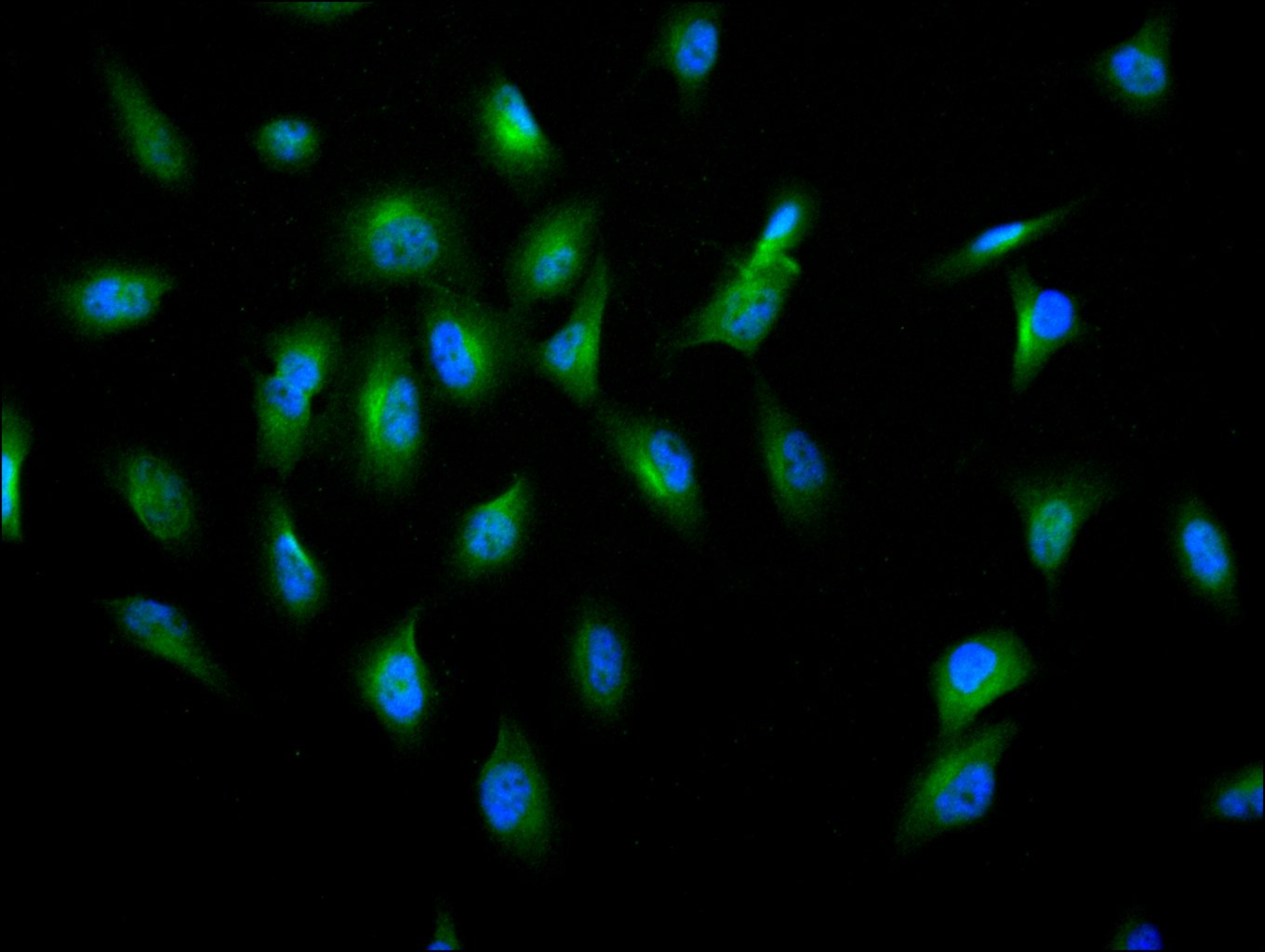
Immunofluorescence staining of U251 cell with CSB-RA555745A0HU at 1:10, counter-stained with DAPI. The cells were fixed in 4% formaldehyde and and permeated by 0.2% TritonX-100 for 15 min. Then 10% normal goat serum to block non-specific protein-protein interactions . The cells were then incubated with the antibody overnight at 4°C. The secondary antibody was Alexa Fluor 488-congugated AffiniPure Goat Anti-Rabbit IgG(H+L) . |
|
|
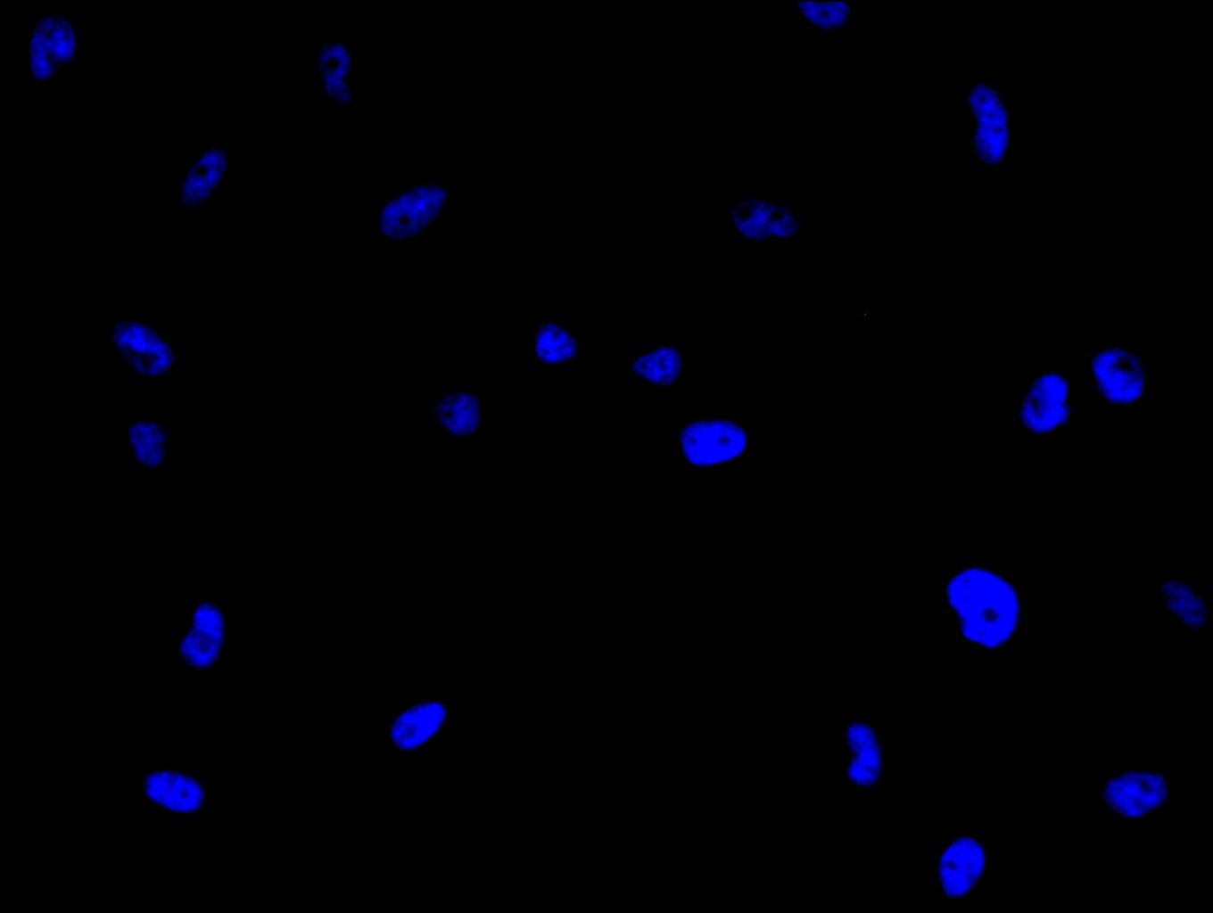
Immunofluorescence staining of U251 cell with 5% goat serum, counter-stained with DAPI. The cells were fixed in 4% formaldehyde and blocked in 10% normal Goat Serum. The cells were then incubated with the antibody overnight at 4C. The secondary antibody was Alexa Fluor 488-congugated AffiniPure Goat Anti-Rabbit IgG(H+L) . |
|
|
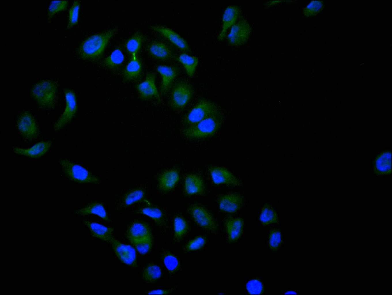
Immunofluorescence staining of Hela cell with CSB-RA555745A0HU at 1:10, counter-stained with DAPI. The cells were fixed in 4% formaldehyde and and permeated by 0.2% TritonX-100 for 15 min. Then 10% normal goat serum to block non-specific protein-protein interactions . The cells were then incubated with the antibody overnight at 4°C. The secondary antibody was Alexa Fluor 488-congugated AffiniPure Goat Anti-Rabbit IgG(H+L) . |
|
|
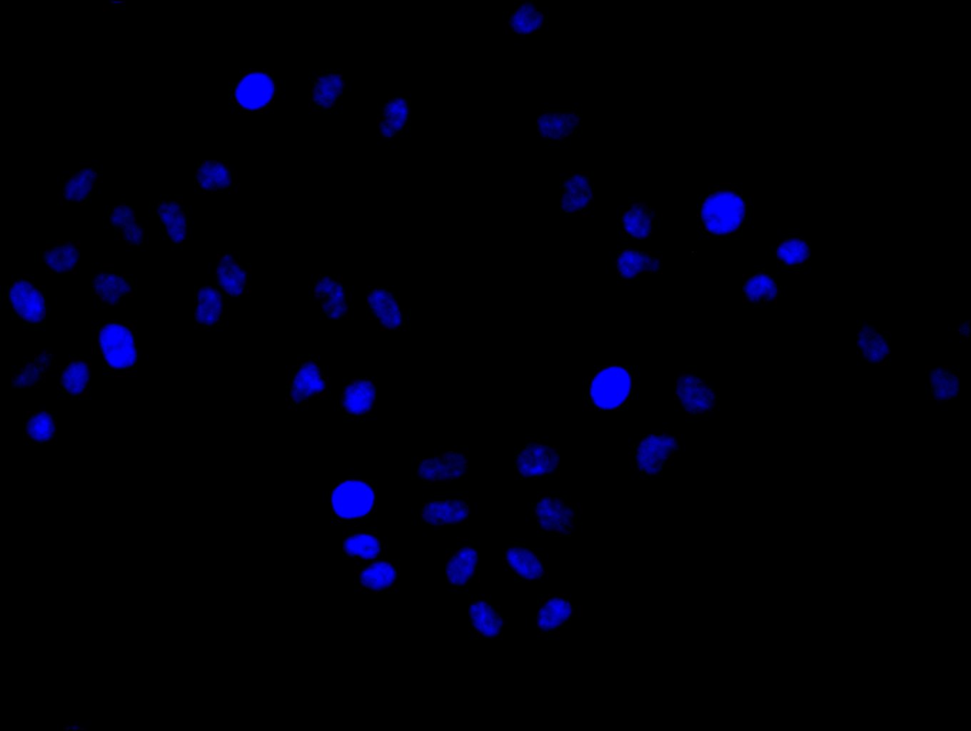
Immunofluorescence staining of Hela cell with 5% goat serum, counter-stained with DAPI. The cells were fixed in 4% formaldehyde and blocked in 10% normal Goat Serum. The cells were then incubated with the antibody overnight at 4C. The secondary antibody was Alexa Fluor 488-congugated AffiniPure Goat Anti-Rabbit IgG(H+L) . |
|
|
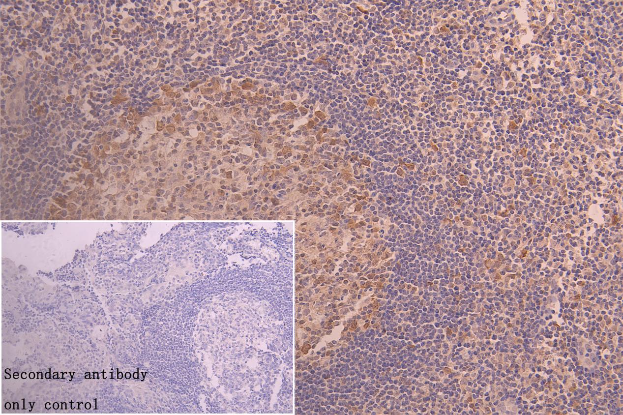
IHC image of CSB-RA555745A0HU diluted at 1:50 and staining in paraffin-embedded human tonsil tissue performed on a Leica BondTM system. After dewaxing and hydration, antigen retrieval was mediated by high pressure in a citrate buffer (pH 6.0) . Section was blocked with 10% normal goat serum 30min at RT. Then primary antibody (1% BSA) was incubated at 4C overnight. The primary is detected by a Goat anti-rabbit polymer IgG labeled by HRP and visualized using 0.05% DAB. |
|
|
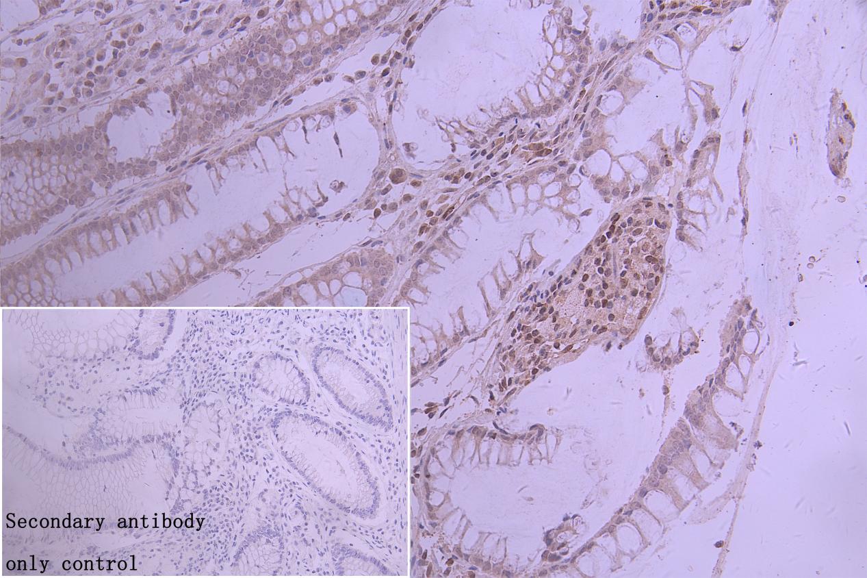
IHC image of CSB-RA555745A0HU diluted at 1:50 and staining in paraffin-embedded human colorectal cancer performed on a Leica BondTM system. After dewaxing and hydration, antigen retrieval was mediated by high pressure in a citrate buffer (pH 6.0) . Section was blocked with 10% normal goat serum 30min at RT. Then primary antibody (1% BSA) was incubated at 4C overnight. The primary is detected by a Goat anti-rabbit polymer IgG labeled by HRP and visualized using 0.05% DAB.Secondary antibody only control: uses 1% BSA instead of primary antibody |
|
|
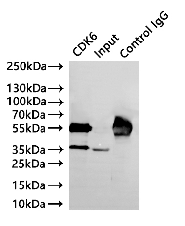
Immunoprecipitating CDK6 in K562whole cell lysate Lane 1: CSB-RA555745A0HU(3µg) + K562 whole cell lysate(220µg) Lane 2: K562 whole cell lysate(30µg) Lane 3:Rabbit control IgG instead of CSB-RA010605A0HUinK562wholecelllysate For western blotting, a HRP-conjugated Protein G antibody was used as the secondary antibody (1/40000) |
|
|
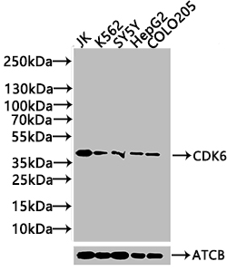
Western Blot Positive WB detected in: JK whole cell lysate(20µg) , K562 whole cell lysate(20µg) , SY5Y whole cell lysate(20µg) , HepG2 whole cell lysate(20µg) , COLO205 whole cell lysate(20µg) All lanes: CDK6 antibody at 1:1000 Secondary Goat polyclonal to rabbit IgG at 1/40000 dilution Predicted band size: 37 kDa Observed band size: 37 kDa Exposure time: 120s |
Produktgarantie und fachkundiger Support
