
YWHAB Recombinant Monoclonal Antibody, Clone: [6F6], Unconjugated, Rabbit
Artikelnummer:
CSB-RA616077A0HU
- Bilder (8)
| Artikelname: | YWHAB Recombinant Monoclonal Antibody, Clone: [6F6], Unconjugated, Rabbit |
| Artikelnummer: | CSB-RA616077A0HU |
| Hersteller Artikelnummer: | CSB-RA616077A0HU |
| Alternativnummer: | CSB-RA616077A0HU-100UL, CSB-RA616077A0HU-50UL |
| Hersteller: | Cusabio |
| Wirt: | Rabbit |
| Kategorie: | Antikörper |
| Applikation: | ELISA, FC, IF, IHC, WB |
| Spezies Reaktivität: | Human, Mouse, Rat |
| Konjugation: | Unconjugated |
| Alternative Synonym: | 14-3-3 protein beta/alpha (Protein 1054) (Protein kinase C inhibitor protein 1) (KCIP-1) [Cleaved into: 14-3-3 protein beta/alpha, N-terminally processed], YWHAB |
| Klonalität: | Monoclonal |
| Klon-Bezeichnung: | [6F6] |
| UniProt: | P31946 |
| Puffer: | Rabbit IgG in 10mM phosphate buffered saline , pH 7.4, 150mM sodium chloride, 0.05% BSA, 0.02% sodium azide and 50% glycerol. |
| Reinheit: | Affinity-chromatography |
| Formulierung: | Liquid |
| Target-Kategorie: | YWHAB |
| Antibody Type: | Recombinant Antibody |
| Application Verdünnung: | Recommended dilution: WB:1:500-1:2000, IHC:1:50-1:200 IF:1:20-1:100, FC:1:50-1:200 |
|
|
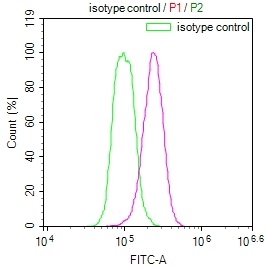
Overlay Peak curve showing Hela cells stained with CSB-RA616077A0HU (red line) at 1:100. The cells were fixed in 4% formaldehyde (15min) and permeated by 0.2% TritonX-100 for 10min. Then 10% normal goat serum to block non-specific protein-protein interactions followed by the antibody (1ug/1*106cells) for 45min at 4°C. The secondary antibody used was FITC-conjugated Goat Anti-rabbit IgG(H+L) at 1:200 dilution for 35min at 4°C.Control antibody (green line) was rabbit IgG (1ug/1*106cells) used under the same conditions. Acquisition of >10,000 events was performed. |
|
|
|
|
|
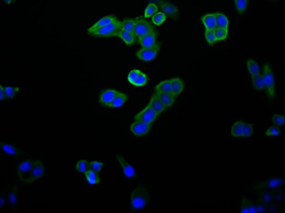
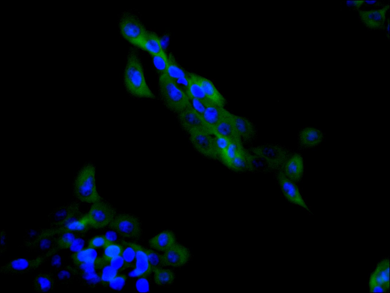
Immunofluorescence staining of A431 cell with CSB-RA616077A0HU at 1:20, counter-stained with DAPI. The cells were fixed in 4% formaldehyde and and permeated by 0.2% TritonX-100 for 15 min. Then 10% normal goat serum to block non-specific protein-protein interactions . The cells were then incubated with the antibody overnight at 4°C. The secondary antibody was Alexa Fluor 488-congugated AffiniPure Goat Anti-Rabbit IgG(H+L) . |
|
|
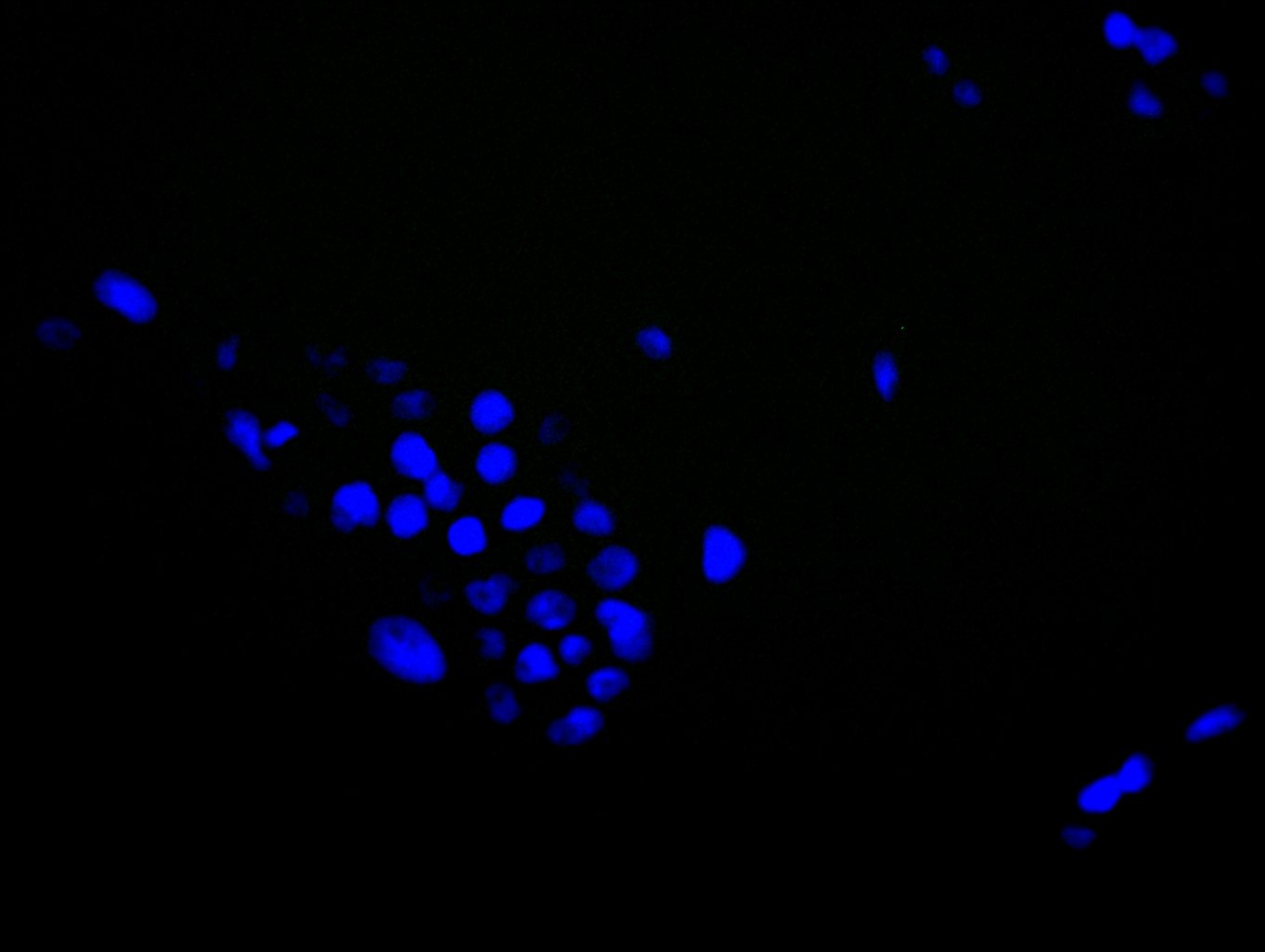
Immunofluorescence staining of A431 cell with 5% goat serum, counter-stained with DAPI. The cells were fixed in 4% formaldehyde and blocked in 10% normal Goat Serum. The cells were then incubated with the antibody overnight at 4C. The secondary antibody was Alexa Fluor 488-congugated AffiniPure Goat Anti-Rabbit IgG(H+L) . |
|
|
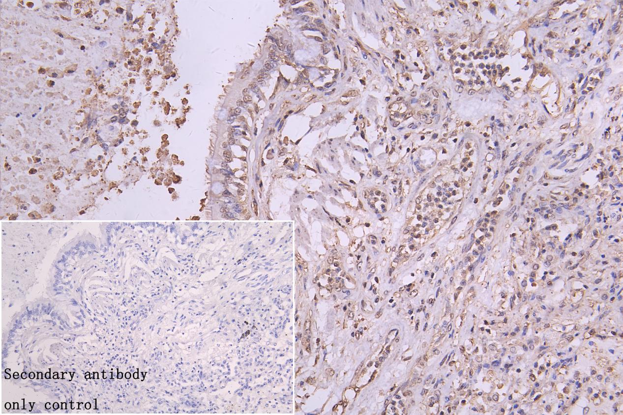
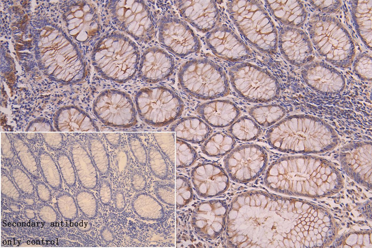
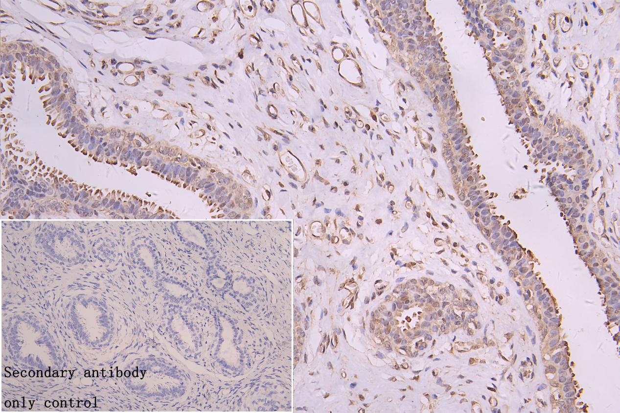
IHC image of CSB-RA616077A0HU diluted at 1:50 and staining in paraffin-embedded human lung cancer performed on a Leica BondTM system. After dewaxing and hydration, antigen retrieval was mediated by high pressure in a citrate buffer (pH 6.0) . Section was blocked with 10% normal goat serum 30min at RT. Then primary antibody (1% BSA) was incubated at 4C overnight. The primary is detected by a Goat anti-rabbit polymer IgG labeled by HRP and visualized using 0.05% DAB. Secondary antibody only control: uses 1% BSA instead of primary antibody |
|
|
IHC image of CSB-RA616077A0HU diluted at 1:50 and staining in paraffin-embedded human colorectal cancer performed on a Leica BondTM system. After dewaxing and hydration, antigen retrieval was mediated by high pressure in a citrate buffer (pH 6.0) . Section was blocked with 10% normal goat serum 30min at RT. Then primary antibody (1% BSA) was incubated at 4C overnight. The primary is detected by a Goat anti-rabbit polymer IgG labeled by HRP and visualized using 0.05% DAB. Secondary antibody only control: uses 1% BSA instead of primary antibody |
|
|
IHC image of CSB-RA616077A0HU diluted at 1:50 and staining in paraffin-embedded human breast cancer performed on a Leica BondTM system. After dewaxing and hydration, antigen retrieval was mediated by high pressure in a citrate buffer (pH 6.0) . Section was blocked with 10% normal goat serum 30min at RT. Then primary antibody (1% BSA) was incubated at 4C overnight. The primary is detected by a Goat anti-rabbit polymer IgG labeled by HRP and visualized using 0.05% DAB.Secondary antibody only control: uses 1% BSA instead of primary antibody |
|
|
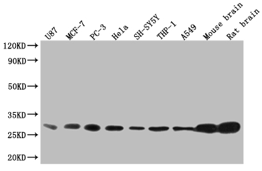
Western Blot Positive WB detected in: U87 whole cell lysate, MCF-7 whole cell lysate, PC3 whole cell lysate, Hela whole cell lysate, SH-SY5Y whole cell lysate, THP-1 whole cell lysate, A549 whole cell lysate, Mouse brain tissue, Rat brain tissue All lanes: YWHAB antibody at 1:2000 Secondary Goat polyclonal to rabbit IgG at 1/50000 dilution Predicted band size: 29, 28 kDa Observed band size: 25-35 kDa |
Produktgarantie und fachkundiger Support
