
Anti-GFAP (Hu) from Mouse (Clone: IF3)
Artikelnummer:
DNA-DIA-700-P05
- Bilder (2)
| Artikelname: | Anti-GFAP (Hu) from Mouse (Clone: IF3) |
| Artikelnummer: | DNA-DIA-700-P05 |
| Hersteller Artikelnummer: | DNA-DIA-700-P05 |
| Alternativnummer: | DNA-DIA-700-P05 |
| Hersteller: | dianova |
| Wirt: | Mouse |
| Kategorie: | Antikörper |
| Applikation: | IHC |
| Spezies Reaktivität: | Human |
| Immunogen: | Fusionsprotein |
| Konjugation: | Unconjugated |
| Alternative Synonym: | Glial fibrillary acidic protein,GFAP,gfapl,Intermediate Filament Protein |
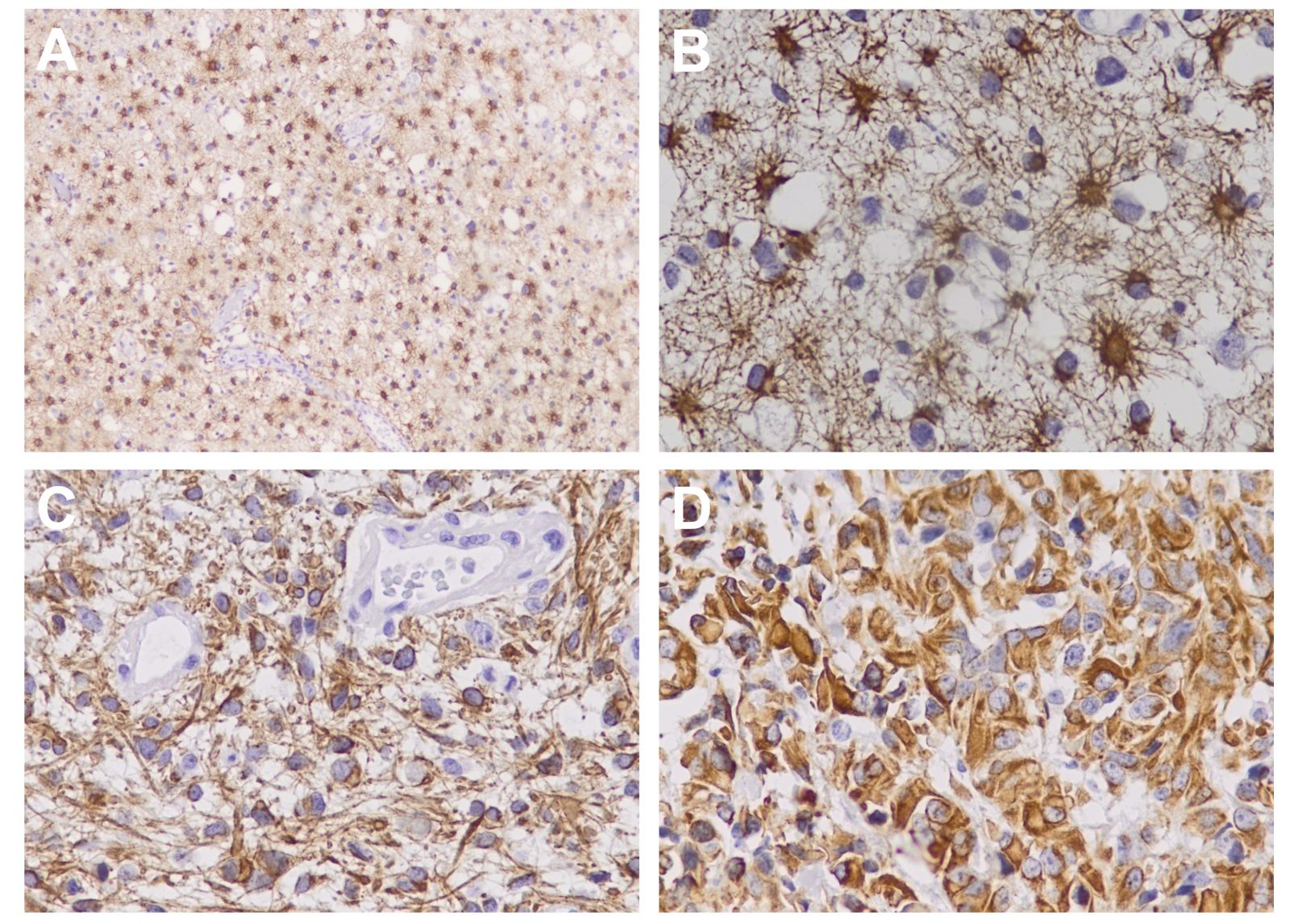
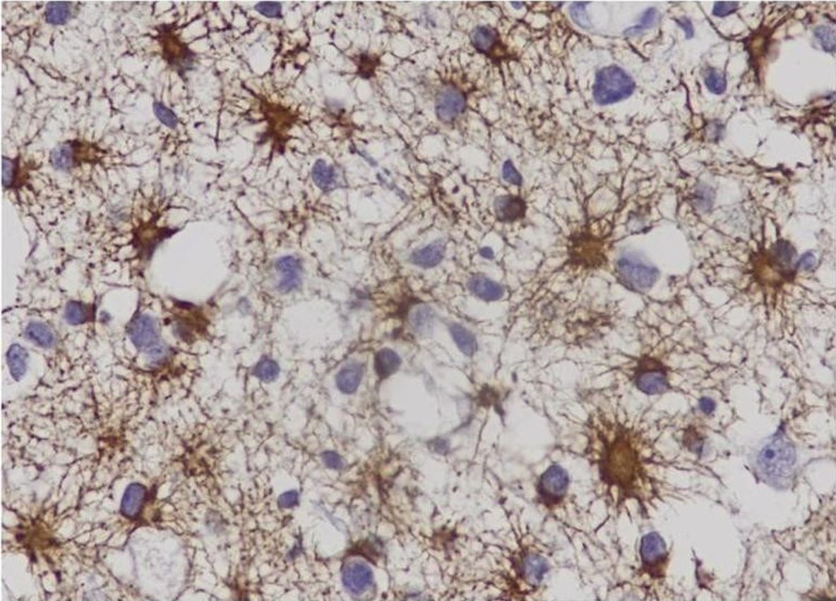
| Anti-GFAP (Hu) aus Maus (Klon: IF3), IgG1, Unconjugated, Mouse, Monoclonal GFAP (Glial Fibrillary Acidic Protein) has proved to be the most specific marker for cells of astrocytic origin that distinguishes differentiated astrocytes from other glial cells during the development of the central nervous system. Monoclonal antibodies to GFAP are useful in differentiating primary gliomas from metastatic lesions in the brain and for documenting astrocytic differentia-tion in tumors outside the CNS. As a 50 kDa intracytoplasmic filamentous protein, GFAP is thought to be important in astrocyte motility and shape by providing structural stability. As a consequence of injury to the human CNS caused by trauma, genetic disorders, or chemicals, GFAP is markly upregulated and astrocytes proliferate. On the other hand, with increasing astrocyte malignancy, a progressive loss of GFAP production has been observed. Consequntly, malignant astrocytomas have fewer tumour cells that stain positively and in-tensely for GFAP than do less malignant astrocytomas and normal brain specimens. Outside the CNS, GFAP has been demon-strated in Schwann cells, enteric glia cells, salivary gland neoplasms, and metastasizing renal carcinomas. Moreover, GFAP has been detected in epiglottic cartilage, pituicytes, immature oligodendrocytes, papillary meningiomas, and myoepithelial cells of the breast. |
| Application Verdünnung: | Histo-/Zytochemie 1:300 - 1:600 |
|
|
Abb1_IHCA-D_GFAP_DIA-700 |
|
|
Asterocytoma-grade-II-40x |
Produktgarantie und fachkundiger Support
