AP-1 rabbit pAb, Rabbit, Polyclonal
Artikelnummer:
EBT-ES1668
Produktübersicht
Artikelname:
AP-1 rabbit pAb, Rabbit, Polyclonal
Artikelnummer:
EBT-ES1668
Hersteller Artikelnummer:
ES1668
Alternativnummer:
EBT-ES1668-50, EBT-ES1668-100
Hersteller:
ELK Biotechnology
Wirt:
Rabbit
Kategorie:
Antikörper
Applikation:
ELISA, IF, IHC, IP, WB
Spezies Reaktivität:
Human, Mouse, Rat
Immunogen:
The antiserum was produced against synthesized peptide derived from human c-Jun. AA range:58-107
Alternative Synonym:
JUN, Transcription factor AP-1, Activator protein 1, AP1, Proto-oncogene c-Jun, V-jun avian sarcoma virus 17 oncogene homolog, p39
Produktbeschreibung
This gene is the putative transforming gene of avian sarcoma virus 17. It encodes a protein which is highly similar to the viral protein, and which interacts directly with specific target DNA sequences to regulate gene expression. This gene is intronless and is mapped to 1p32-p31, a chromosomal region involved in both translocations and deletions in human malignancies. [provided by RefSeq, Jul 2008],
Produkteigenschaften
Klonalität:
Polyclonal
Konzentration:
1 mg/ml
Molekulargewicht:
39-42kD
NCBI:
3725
UniProt:
P05412
Anwendungsbeschreibung
Application Verdünnung:
IF: 1:50-200 Western Blot: 1/500 - 1/2000. Immunohistochemistry: 1/100 - 1/300. Immunoprecipitation: 2-5 ug/mg lysate. ELISA: 1/20000. Not yet tested in other applications.
Bilder
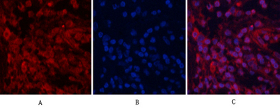
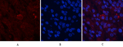
Immunofluorescence analysis of human-lung tissue. 1,AP-1 Polyclonal Antibody(red) was diluted at 1:200(4C,overnight). 2, Cy3 labled Secondary antibody was diluted at 1:300(room temperature, 50min).3, Picture B: DAPI(blue) 10min. Picture A:Target. Picture B: DAPI. Picture C: merge of A+B
Immunofluorescence analysis of human-lung tissue. 1,AP-1 Polyclonal Antibody(red) was diluted at 1:200(4C,overnight). 2, Cy3 labled Secondary antibody was diluted at 1:300(room temperature, 50min).3, Picture B: DAPI(blue) 10min. Picture A:Target. Picture B: DAPI. Picture C: merge of A+B
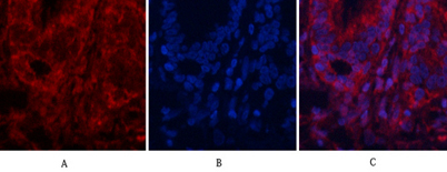
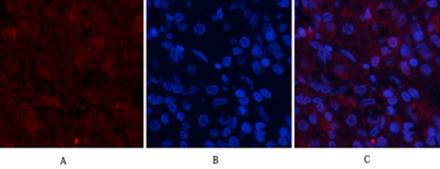
Immunofluorescence analysis of human-stomach tissue. 1,AP-1 Polyclonal Antibody(red) was diluted at 1:200(4C,overnight). 2, Cy3 labled Secondary antibody was diluted at 1:300(room temperature, 50min).3, Picture B: DAPI(blue) 10min. Picture A:Target. Picture B: DAPI. Picture C: merge of A+B
Immunofluorescence analysis of human-stomach tissue. 1,AP-1 Polyclonal Antibody(red) was diluted at 1:200(4C,overnight). 2, Cy3 labled Secondary antibody was diluted at 1:300(room temperature, 50min).3, Picture B: DAPI(blue) 10min. Picture A:Target. Picture B: DAPI. Picture C: merge of A+B