Cdc2 rabbit pAb, Rabbit, Polyclonal

Artikelnummer: EBT-ES1922
Artikelname: Cdc2 rabbit pAb, Rabbit, Polyclonal
Artikelnummer: EBT-ES1922
Hersteller Artikelnummer: ES1922
Alternativnummer: EBT-ES1922-50, EBT-ES1922-100
Hersteller: ELK Biotechnology
Wirt: Rabbit
Kategorie: Antikörper
Applikation: ELISA, IF, IHC, WB
Spezies Reaktivität: Human, Mouse, Rat
Immunogen: 
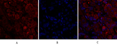
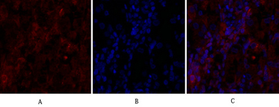
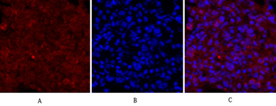
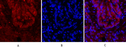
The antiserum was produced against synthesized peptide derived from human CDC2. AA range:5-54
Alternative Synonym: CDK1, CDC2, CDC28A, CDKN1, P34CDC2, Cyclin-dependent kinase 1, CDK1, Cell division control protein 2 homolog, Cell division protein kinase 1, p34 protein kinase
cyclin dependent kinase 1(CDK1) Homo sapiens The protein encoded by this gene is a member of the Ser/Thr protein kinase family. This protein is a catalytic subunit of the highly conserved protein kinase complex known as M-phase promoting factor (MPF), which is essential for G1/S and G2/M phase transitions of eukaryotic cell cycle. Mitotic cyclins stably associate with this protein and function as regulatory subunits. The kinase activity of this protein is controlled by cyclin accumulation and destruction through the cell cycle. The phosphorylation and dephosphorylation of this protein also play important regulatory roles in cell cycle control. Alternatively spliced transcript variants encoding different isoforms have been found for this gene. [provided by RefSeq, Mar 2009],
Klonalität: Polyclonal
Konzentration: 1 mg/ml
Molekulargewicht: 34kD
NCBI: 983
UniProt: P06493
Application Verdünnung: IF: 1:50-200 Western Blot: 1/500 - 1/2000. ELISA: 1/10000. Not yet tested in other applications.
Immunofluorescence analysis of human-stomach tissue. 1,Cdc2 Polyclonal Antibody(red) was diluted at 1:200(4C,overnight). 2, Cy3 labled Secondary antibody was diluted at 1:300(room temperature, 50min).3, Picture B: DAPI(blue) 10min. Picture A:Target. Picture B: DAPI. Picture C: merge of A+B
Immunofluorescence analysis of rat-lung tissue. 1,Cdc2 Polyclonal Antibody(red) was diluted at 1:200(4C,overnight). 2, Cy3 labled Secondary antibody was diluted at 1:300(room temperature, 50min).3, Picture B: DAPI(blue) 10min. Picture A:Target. Picture B: DAPI. Picture C: merge of A+B