
CD2AP antibody, Unconjugated, Rabbit, Polyclonal
Artikelnummer:
GTX106235
- Bilder (10)
| Artikelname: | CD2AP antibody, Unconjugated, Rabbit, Polyclonal |
| Artikelnummer: | GTX106235 |
| Hersteller Artikelnummer: | GTX106235 |
| Alternativnummer: | GTX106235-100,GTX106235-25 |
| Hersteller: | GeneTex |
| Wirt: | Rabbit |
| Kategorie: | Antikörper |
| Applikation: | ICC, IHC-P, WB |
| Spezies Reaktivität: | Human, Mouse, Rat |
| Immunogen: | Recombinant protein encompassing a sequence within the center region of human CD2AP. The exact sequence is proprietary. |
| Konjugation: | Unconjugated |
| Alternative Synonym: | CD2 associated protein , CMS |
| Klonalität: | Polyclonal |
| Konzentration: | 0.17 mg/ml (Please refer to the vial label for the specific concentration.) |
| Molekulargewicht: | 71 |
| NCBI: | 23607 |
| UniProt: | Q9Y5K6 |
| Puffer: | 1XPBS (pH7), 1% BSA, 20% Glycerol, 0.025% ProClin 300. |
| Reinheit: | Purified by antigen-affinity chromatography. |
| Formulierung: | Liquid |
| Anwendungsbeschreibung: | WB: 1:500-1:3000. ICC/IF: 1:100-1:1000. IHC-P: 1:100-1:1000. *Optimal dilutions/concentrations should be determined by the researcher.Not tested in other applications. |
|
|
GTX106235 WB Image |
|
|
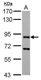
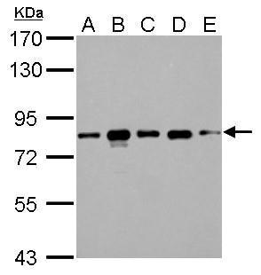
CD2AP antibody detects CD2AP protein by western blot analysis. A. 30 μg 293T whole cell lysate/extract B. 30 μg A431 whole cell lysate/extract C. 30 μg HeLa whole cell lysate/extract D. 30 μg HepG2 whole cell lysate/extract E. 30 μg A375 whole cell lysate/extract 7.5% SDS-PAGE CD2AP antibody (GTX106235) dilution: 1:1000 The HRP-conjugated anti-rabbit IgG antibody (GTX213110-01) was used to detect the primary antibody. |
|
|
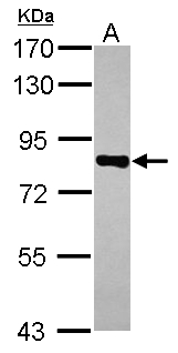
Sample (30 ug of whole cell lysate) A: A431 7.5% SDS PAGE GTX106235 diluted at 1:1000 |
|
|
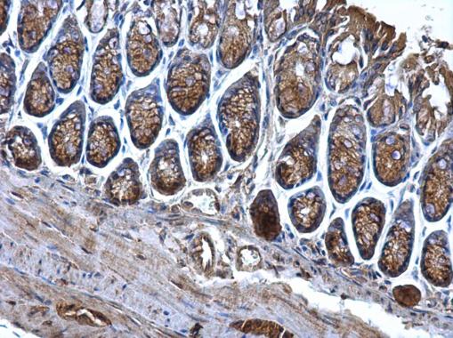
CD2AP antibody detects CD2AP protein at cytosol on mouse colon by immunohistochemical analysis. Sample: Paraffin-embedded mouse colon. CD2AP antibody (GTX106235) dilution: 1:500. Antigen Retrieval: Trilogy™ (EDTA based, pH 8.0) buffer, 15min |
|
|
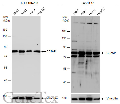
Various whole cell extracts (30 μg) were separated by 7.5% SDS-PAGE, and the membranes were blotted with CD2AP antibody (GTX106235) diluted at 1:1000 and competitor's antibody (sc-9137) diluted at 1:200. The HRP-conjugated anti-rabbit IgG antibody (GTX213110-01) was used to detect the primary antibody. *The competitor is not affiliated with GeneTex and does not endorse this product. |
|
|
Confocal immunofluorescence analysis (Olympus FV10i) of paraformaldehyde-fixed A431, using CD2AP(GTX106235) antibody (Green) at 1:500 dilution. Alpha-tubulin filaments were labeled with GTX11304 (Red) at 1:500. |
|
|
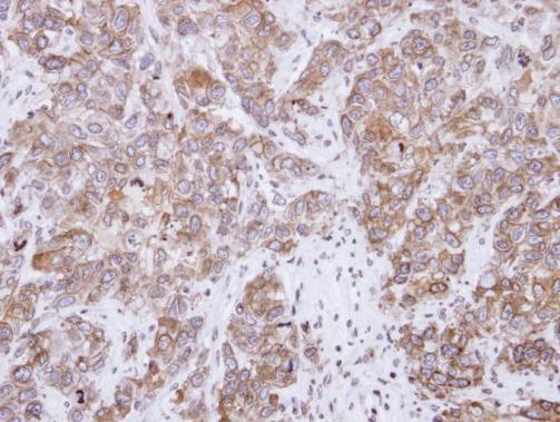
Immunohistochemical analysis of paraffin-embedded FaDu xenograft, using CD2-associated protein(GTX106235) antibody at 1:500 dilution. Antigen Retrieval: Trilogy™ (EDTA based, pH 8.0) buffer, 15min |
|
|
Various whole cell extracts (30 μg) were separated by 7.5% SDS-PAGE, and the membranes were blotted with CD2AP antibody (GTX106235) diluted at 1:1000 and competitor's antibody (sc-9137) diluted at 1:200. The HRP-conjugated anti-rabbit IgG antibody (GTX213110-01) was used to detect the primary antibody. |
|
|
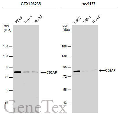
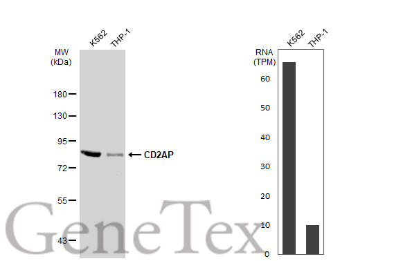
Various whole cell extracts (30 μg) were separated by 7.5% SDS-PAGE, and the membrane was blotted with CD2AP antibody (GTX106235) diluted at 1:1000. The HRP-conjugated anti-rabbit IgG antibody (GTX213110-01) was used to detect the primary antibody. Corresponding RNA expression data for the same cell lines are based on Human Protein Atlas program. |
|
|
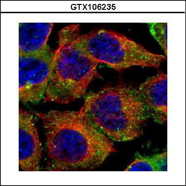
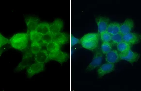
CD2AP antibody detects CD2AP protein at cell membrane and cytoplasm by immunofluorescent analysis.Sample: A431 cells were fixed in ice-cold MeOH for 5 min.Green: CD2AP stained by CD2AP antibody (GTX106235) diluted at 1:500.Blue: Fluoroshield with DAPI (GTX30920). |
Produktgarantie und fachkundiger Support
