
SLC25A13 antibody [N3C2], Internal, Unconjugated, Rabbit, Polyclonal
Artikelnummer:
GTX109001
- Bilder (9)
| Artikelname: | SLC25A13 antibody [N3C2], Internal, Unconjugated, Rabbit, Polyclonal |
| Artikelnummer: | GTX109001 |
| Hersteller Artikelnummer: | GTX109001 |
| Alternativnummer: | GTX109001-100,GTX109001-25 |
| Hersteller: | GeneTex |
| Wirt: | Rabbit |
| Kategorie: | Antikörper |
| Applikation: | ICC, IHC-P, WB |
| Spezies Reaktivität: | Human, Mouse, Rat |
| Immunogen: | Recombinant protein encompassing a sequence within the center region of human SLC25A13. The exact sequence is proprietary. |
| Konjugation: | Unconjugated |
| Alternative Synonym: | solute carrier family 25 member 13 , ARALAR2 , CITRIN , CTLN2 |
| Anwendungsbeschreibung: | WB: 1:500-1:3000. ICC/IF: 1:100-1:1000. IHC-P: 1:100-1:1000. *Optimal dilutions/concentrations should be determined by the researcher.Not tested in other applications. |
|
|
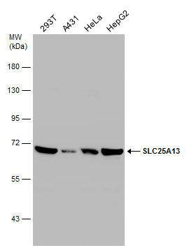
GTX109001 WB Image |
|
|
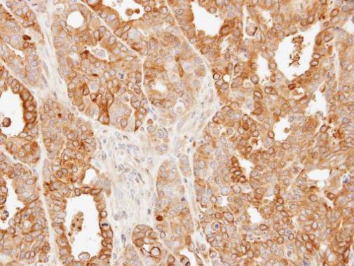
Immunohistochemical analysis of paraffin-embedded N87 xenograft, using SLC25A13(GTX109001) antibody at 1:500 dilution. Antigen Retrieval: Trilogy™ (EDTA based, pH 8.0) buffer, 15min |
|
|
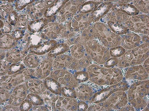
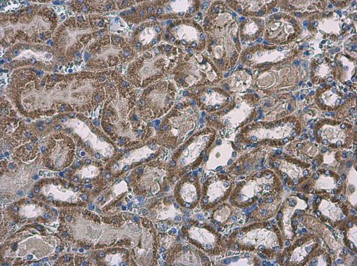
SLC25A13 antibody [N3C2], Internal detects SLC25A13 protein at cytoplasm in mouse kidney by immunohistochemical analysis. Sample: Paraffin-embedded mouse kidney. SLC25A13 antibody [N3C2], Internal (GTX109001) diluted at 1:500. Antigen Retrieval: Citrate buffer, pH 6.0, 15 min |
|
|
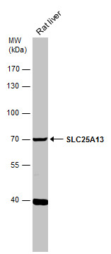
Rat tissue extract (50 μg) was separated by 7.5% SDS-PAGE, and the membrane was blotted with SLC25A13 antibody [N3C2], Internal (GTX109001) diluted at 1:500. The HRP-conjugated anti-rabbit IgG antibody (GTX213110-01) was used to detect the primary antibody. |
|
|
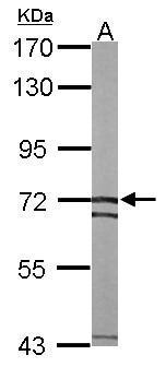
Sample (50 μg of whole cell lysate) A: mouse liver 7.5% SDS PAGE GTX109001 diluted at 1:1000 The HRP-conjugated anti-rabbit IgG antibody (GTX213110-01) was used to detect the primary antibody. |
|
|
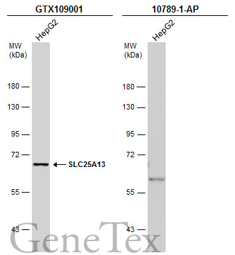
Whole cell extract (30 μg) was separated by 7.5% SDS-PAGE, and the membranes were blotted with SLC25A13 antibody [N3C2], Internal (GTX109001) diluted at 1:1000 and competitor's antibody (10789-1-AP) diluted at 1:1000. The HRP-conjugated anti-rabbit IgG antibody (GTX213110-01) was used to detect the primary antibody. *The competitor is not affiliated with GeneTex and does not endorse this product. |
|
|
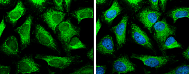
SLC25A13 antibody [N3C2], Internal detects SLC25A13 protein at mitochondria by immunofluorescent analysis. Sample: HeLa cells were fixed in 4% paraformaldehyde at RT for 15 min. Green: SLC25A13 protein stained by SLC25A13 antibody [N3C2], Internal (GTX109001) diluted at 1:500. Blue: Hoechst 33342 staining. |
|
|
SLC25A13 antibody [N3C2], Internal detects SLC25A13 protein at cytoplasm in mouse kidney by immunohistochemical analysis. Sample: Paraffin-embedded mouse kidney. SLC25A13 antibody [N3C2], Internal (GTX109001) diluted at 1:500. Antigen Retrieval: Citrate buffer, pH 6.0, 15 min |
|
|
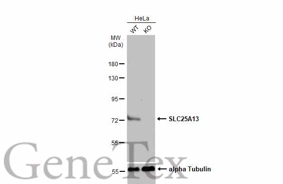
Wild-type (WT) and SLC25A13 knockout (KO) HeLa cell extracts (30 μg) were separated by 7.5% SDS-PAGE, and the membrane was blotted with SLC25A13 antibody [N3C2], Internal (GTX109001) diluted at 1:3000. The HRP-conjugated anti-rabbit IgG antibody (GTX213110-01) was used to detect the primary antibody. |
Produktgarantie und fachkundiger Support
