p53 Tumor Suppressor Protein Antibody, IgG2b, Clone: [SPM590], Mouse, Monoclonal
Artikelnummer:
NBT-7157-MSM2X-P1ABX
Hersteller Artikelnummer:
7157-MSM2X-P1ABX
Alternativnummer:
NBT-7157-MSM2X-P1ABX-100
Hersteller:
NeoBiotechnologies
Wirt:
Mouse
Kategorie:
Antikörper
Applikation:
FC, IF, IHC, WB
Spezies Reaktivität:
Bovine, Human, Monkey
Immunogen:
Recombinant human wild type p53 protein expressed in E. coli.
Alternative Synonym:
Antigen NY-CO-13, BCC7, Cellular Tumor Antigen p53, LFS1, TP53, Transformation Related Protein 53 (TRP53), Tumor Protein p53, Tumor Suppressor p53
Recognizes a 53kDa protein, which is identified as p53 suppressor gene product. It reacts with the mutant as well as the wild form of p53. Its epitope maps within the N-terminus (aa 37-45) of p53. p53 is a tumor suppressor gene expressed in a wide variety of tissue types and is involved in regulating cell growth, replication, and apoptosis. It binds to MDM2, SV40 T antigen and human papilloma virus E6 protein. Positive nuclear staining with p53 antibody has been reported to be a negative prognostic factor in breast carcinoma, lung carcinoma, colorectal, and urothelial carcinoma. Anti-p53 positivity has also been used to differentiate uterine serous carcinoma from endometrioid carcinoma as well as to detect intratubular germ cell neoplasia. Mutations involving p53 are found in a wide variety of malignant tumors, including breast, ovarian, bladder, colon, lung, and melanoma.
200ug/ml of Ab Purified from Bioreactor Concentrate by Protein A/G. Prepared in 10mM PBS with 0.05% BSA & 0.05% azide. Also available WITHOUT BSA & azide at 1.0mg/ml.
Antibody Type:
Monoclonal Antibody
Anwendungsbeschreibung:
Flow Cytometry (1-2ug/million cells), Immunofluorescence (1-2ug/ml), Western Blot (1-2ug/ml), Immunohistochemistry (Formalin-fixed) (1-2ug/ml for 30 minutes at RT) (Staining of formalin-fixed tissues requires heating tissue sections in 10mM Tris with 1mM
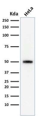
Western Blot Analysis of human HeLa cell lysate using p53 Monoclonal Antibody (SPM590)
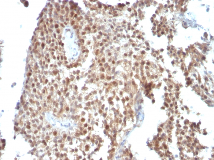
Formalin-fixed, paraffin-embedded human Bladder Carcinoma stained with p53 Monoclonal Antibody (SPM590)
Western Blot Analysis of human HeLa cell lysate using p53 Monoclonal Antibody (SPM590)
* Mehrwertsteuer und Versandkosten nicht enthalten. Irrtümer und Preisänderungen vorbehalten