CD63 is expressed on activated platelets, monocytes and macrophages, and is weakly expressed on granulocytes, T cell and B cells. It is located on the basophilic granule membranes and on the plasma membranes of lymphocytes and granulocytes. CD63 is a member of the TM4 superfamily of leukocyte glycoproteins that includes CD9, CD37 and CD53, which contain four transmembrane regions. CD63 may play a role in phagocytic and intracellular lysosome-phagosome fusion events. CD63 deficiency is associated with Hermansky-Pudlak syndrome and is strongly expressed during the early stages of melanoma progression.
200ug/ml of Ab purified from Bioreactor Concentrate by Protein A/G. Prepared in 10mM PBS with 0.05% BSA & 0.05% azide. Also available WITHOUT BSA & azide at 1.0mg/ml.
Antibody Type:
Monoclonal Antibody
Anwendungsbeschreibung:
ELISA, Functional studies, Immunoprecipitation (1-2ug per 100-500ug of total protein (1ml of cell lysate)), ,Flow Cytometry (1-2ug/million cells), Immunofluorescence (1-2ug/ml), ,Immunohistochemistry (Formalin-fixed) (1-2ug/ml for 30 minutes at RT),(Stai
SDS-PAGE Analysis of Purified CD63 Mouse Monoclonal Antibody (LAMP3/4949). Confirmation of Purity and Integrity of Antibody.
Formalin-fixed, paraffin-embedded human tonsil stained with CD63 Mouse Monoclonal Antibody (LAMP3/4949). Inset: PBS instead of primary antibody, secondary only negative control.
Flow cytometry analysis of bead-bound exosomes derived from MCF-7 cells. Unstained exosomes (gray), exosomes stained with CF568-labeled CD63 Mouse Monoclonal Antibody (LAMP3/4949) (orange).
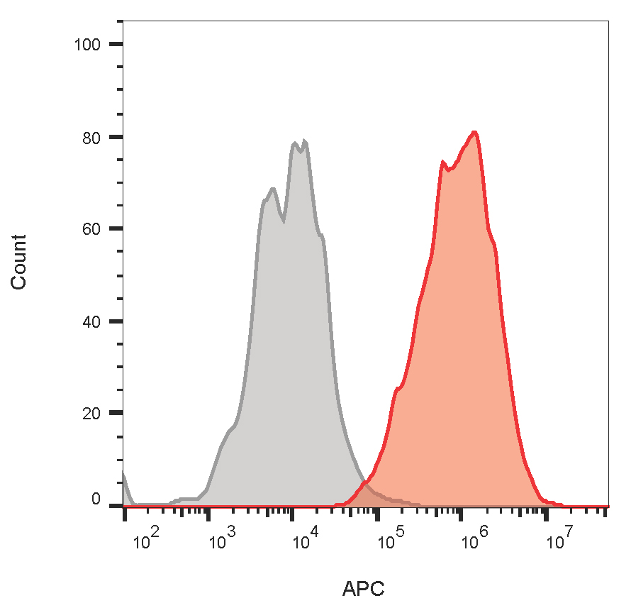
Flow cytometric analysis using MCF-7 cells. CD63 Mouse Monoclonal Antibody (LAMP3/4949) followed by goat anti-mouse IgG-CF640 (red), unstained cells (gray).
* Mehrwertsteuer und Versandkosten nicht enthalten. Irrtümer und Preisänderungen vorbehalten