
CXCR4 Antibody, Unconjugated, Rabbit, Polyclonal
Artikelnummer:
PRS-1012
- Bilder (9)
| Artikelname: | CXCR4 Antibody, Unconjugated, Rabbit, Polyclonal |
| Artikelnummer: | PRS-1012 |
| Hersteller Artikelnummer: | 1012 |
| Alternativnummer: | PRS-1012-0.02,PRS-1012-0.1 |
| Hersteller: | ProSci |
| Wirt: | Rabbit |
| Kategorie: | Antikörper |
| Applikation: | ELISA, IF, IHC, WB |
| Spezies Reaktivität: | Human, Mouse, Rat |
| Immunogen: | Anti-CXCR4 antibody (1012) was raised against a peptide corresponding to 15 amino acids near the center of human CXCR4 isoform b. The immunogen is located within amino acids 170-220 of CXCR4. |
| Konjugation: | Unconjugated |
| Alternative Synonym: | CXCR4 Antibody: FB22, HM89, LAP3, LCR1, NPYR, WHIM, CD184, LESTR, NPY3R, NPYRL, HSY3RR, NPYY3R, D2S201E |
| Application Verdünnung: | Optimal dilutions for each application to be determined by the researcher. |
| Anwendungsbeschreibung: | WB: 1 - 2 µg/mL, IHC-P: 5 µg/mL, IF: 4 µg/mL. Antibody validated: Western Blot in human, mouse, and rat samples, Immunohistochemistry and Immunofluorescence in human samples. All other applications and species not yet tested. |
|
|
Figure 7 KO Validation of CXCR4 by Flow Cytometry (Ödemis, et al., 2010) Astrocytes from wild-type or CXCR4 knockout mice were stained with primary antibodies against CXCR4 and FITC-labeled secondary antibodies, and subsequently subjected to flow cytometry. CXCR4-/- astrocytes (red) showed loss of CXCR4 cell-surface expression compared with wild-type cells (black). |
|
|
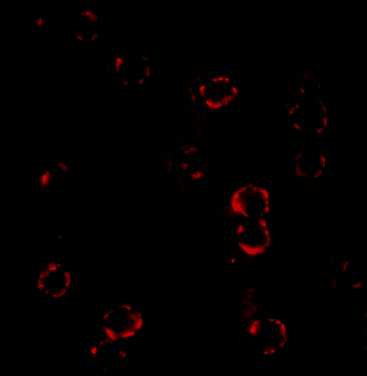
Figure 5 Immunofluorescence Validation of CXCR4 Immunofluorescent analysis of 4% paraformaldehyde-fixed HeLa cells labeling CXCR4 with 1012 at 4 &956,g/mL, followed by goat anti-rabbit IgG secondary antibody at 1/500 dilution (red). Image showing both membrane and cytoplasmic staining on HeLa cell line. |
|
|
Figure 6 Immunohistochemistry Validation of CXCR4 in Human Spleen Immunohistochemical analysis of paraffin-embedded human spleen tissue using anti-CXCR4 antibody (1012) at 5 &956,g/ml. Tissue was fixed with formaldehyde and blocked with 10% serum for 1 h at RT, antigen retrieval was by heat mediation with a citrate buffer (pH6). Samples were incubated with primary antibody overnight at 4&730, C. A goat anti-rabbit IgG H&L (HRP) at 1/250 was used as secondary. Counter stained with Hematoxylin. |
|
|
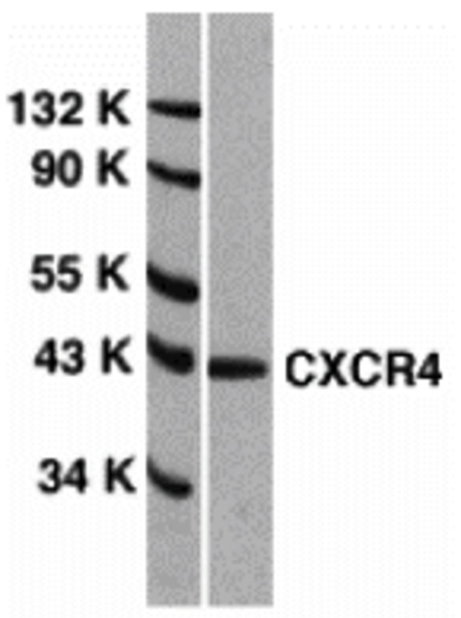
Figure 1 Western Blot Validation of CXCR4 in HeLa Cells Loading: 15 &956,g of lysates per lane.Antibodies: 1012 (1 &956,g/mL), 1 h incubation at RT in 5% NFDM/TBST. Secondary: Goat anti-rabbit IgG HRP conjugate at 1:10000 dilution. |
|
|
Figure 2 Independent Antibody Validation (IAV) via Protein Expression Profile Loading: 15 &956,g of lysates per lane. Antibodies: 1009 (1 &956,g/mL), 1012 (1 &956,g/mL), and beta-actin (1 &956,g/mL), 1 h incubation at RT in 5% NFDM/TBST. Secondary: Goat anti-rabbit IgG HRP conjugate at 1:10000 dilution. |
|
|
Figure 3 Validation with CXCR4 siRNA Knockdown in HeLa Cells HeLa cells were transfected with control siRNAs (lane 1) or CXCR4 siRNAs (lane 2) Loading: 10 &956,g of HeLa whole cell lysates per lane. Antibodies: 1012 (2 &956,g/mL), 1 h incubation at RT in 5% NFDM/TBST. Secondary: Goat anti-rabbit IgG HRP conjugate at 1:10000 dilution. |
|
|
Figure 4 Animal Species Reactivity Loading: Lysates/proteins at 20 &956,g per lane.Antibodies: 1009 (2 &956,g/mL) or 1012 (2 &956,g/mL). 1 h incubation at RT in 5% NFDM/TBST.Secondary: Goat anti-rabbit IgG HRP conjugate at 1:10000 dilution. |
|
|
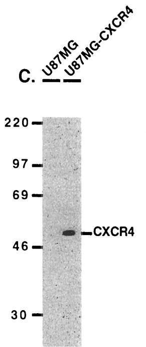
Figure 8 Overexpression Validation of CXCR4 (Kozak et al., 2002) U87MG and U87MG-CXCR4 extracts were included as negative and positive controls, respectively, for CXCR4 detection with anti-CXCR4 antibodies. |
|
|
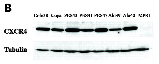
Figure 9 WB Validation of CXCR4 in Human Metastatic Melanoma (Scala et al., 2006) CXCR4 protein was detected in the human metastatic melanoma cell lines and human melanoma cell line (colo38), but not in the human primary melanocytes (MPR1) with anti-CXCR4 antibodies. |
Produktgarantie und fachkundiger Support
