
HSF1 Antibody, Unconjugated, Rabbit, Polyclonal
Artikelnummer:
PRS-15-191
- Bilder (9)
| Artikelname: | HSF1 Antibody, Unconjugated, Rabbit, Polyclonal |
| Artikelnummer: | PRS-15-191 |
| Hersteller Artikelnummer: | 15-191 |
| Alternativnummer: | PRS-15-191-100 |
| Hersteller: | ProSci |
| Wirt: | Rabbit |
| Kategorie: | Antikörper |
| Applikation: | IF, IHC, WB |
| Spezies Reaktivität: | Human, Mouse, Rat |
| Immunogen: | Recombinant fusion protein containing a sequence corresponding to amino acids 350-529 of human HSF1 (NP_005517.1). |
| Konjugation: | Unconjugated |
| Alternative Synonym: | HSF1, HSTF1 |
| Application Verdünnung: | Optimal dilutions for each application to be determined by the researcher. |
| Anwendungsbeschreibung: | WB: 1:500 - 1:2000IHC: 1:50 - 1:100IF: 1:50 - 1:100 |
|
|
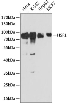
Western blot analysis of extracts of various cell lines, using HSF1 antibody (15-191) at 1:3000 dilution. Secondary antibody: HRP Goat Anti-Rabbit IgG (H+L) at 1:10000 dilution. Lysates/proteins: 25ug per lane. Blocking buffer: 3% nonfat dry milk in TBST. Detection: ECL Basic Kit. Exposure time: 90s. |
|
|
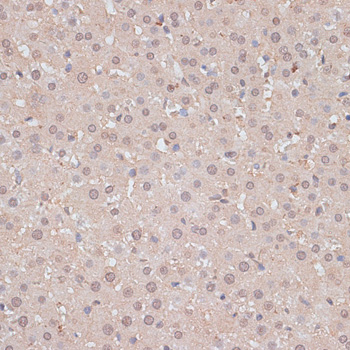
Immunohistochemistry of paraffin-embedded rat liver using HSF1 antibody (15-191) at dilution of 1:100 (40x lens). |
|
|
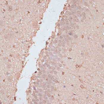
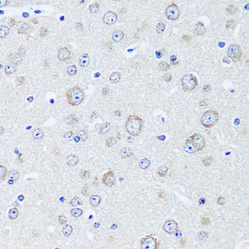
Immunohistochemistry of paraffin-embedded rat brain using HSF1 antibody (15-191) at dilution of 1:100 (40x lens). |
|
|
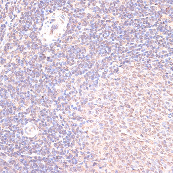
Immunohistochemistry of paraffin-embedded rat spleen using HSF1 antibody (15-191) at dilution of 1:100 (40x lens). |
|
|
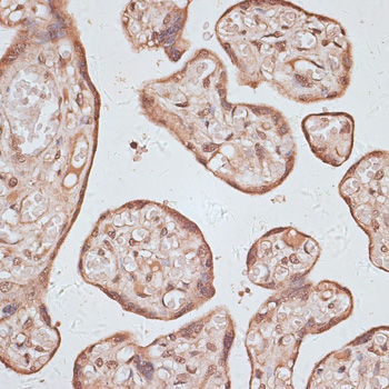
Immunohistochemistry of paraffin-embedded human placenta using HSF1 antibody (15-191) at dilution of 1:100 (40x lens). |
|
|
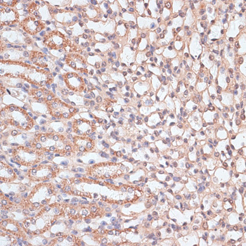
Immunohistochemistry of paraffin-embedded mouse kidney using HSF1 antibody (15-191) at dilution of 1:100 (40x lens). |
|
|
Immunofluorescence analysis of C6 cells using HSF1 antibody (15-191) at dilution of 1:100. Blue: DAPI for nuclear staining. |
|
|
Immunofluorescence analysis of L929 cells using HSF1 antibody (15-191) at dilution of 1:100. Blue: DAPI for nuclear staining. |
|
|
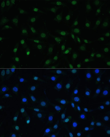
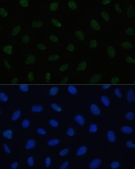
Immunofluorescence analysis of U2OS cells using HSF1 antibody (15-191) at dilution of 1:100. Blue: DAPI for nuclear staining. |
Produktgarantie und fachkundiger Support
