
MonoMethyl-Histone H3-K4 pAb, Unconjugated, Rabbit, Polyclonal
Artikelnummer:
PRS-18-618
- Bilder (8)

| Artikelname: | MonoMethyl-Histone H3-K4 pAb, Unconjugated, Rabbit, Polyclonal |
| Artikelnummer: | PRS-18-618 |
| Hersteller Artikelnummer: | 18-618 |
| Alternativnummer: | PRS-18-618-100 |
| Hersteller: | ProSci |
| Wirt: | Rabbit |
| Kategorie: | Antikörper |
| Applikation: | ChIP, IF, IHC, IP, WB |
| Spezies Reaktivität: | Human, Mouse, Other, Rat |
| Immunogen: | A synthetic methylated peptide corresponding to residues surrounding K4 of human histone H3 |
| Konjugation: | Unconjugated |
| Alternative Synonym: | H3F3A, H3t, H3.4, H3/g, H3FT |
| Application Verdünnung: | Optimal dilutions for each application to be determined by the researcher. |
| Anwendungsbeschreibung: | WB: 1:500 - 1:2000IHC: 1:50 - 1:200IF: 1:50 - 1:200IP: 1:50 - 1:200ChIP: 1:20 - 1:100CHIP seq: 1:20 - 1:100 |
|
|
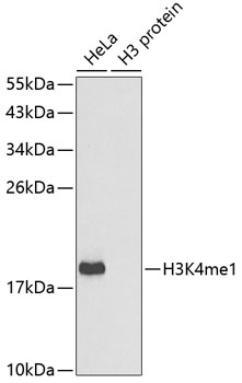
Western blot analysis of extracts of various cell lines, using MonoMethyl-Histone H3-K4 antibody (18-618). Secondary antibody: HRP Goat Anti-Rabbit IgG (H+L) at 1:10000 dilution. Lysates/proteins: 25ug per lane. Blocking buffer: 3% nonfat dry milk in TBST. |
|
|
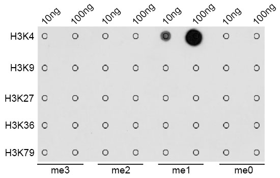
Dot-blot analysis of all sorts of methylation peptides using MonoMethyl-Histone H3-K4 antibody (18-618). |
|
|
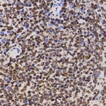
Immunohistochemistry of paraffin-embedded rat spleen using MonoMethyl-Histone H3-K4 antibody (18-618) at dilution of 1:200 (40x lens). |
|
|
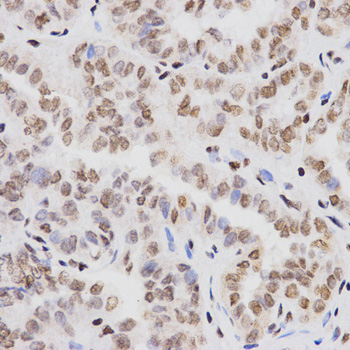
Immunohistochemistry of paraffin-embedded human thyroid cancer using MonoMethyl-Histone H3-K4 antibody (18-618) at dilution of 1:200 (40x lens). |
|
|
Immunohistochemistry of paraffin-embedded rat brain using MonoMethyl-Histone H3-K4 antibody (18-618) at dilution of 1:200 (40x lens). |
|
|
Immunohistochemistry of paraffin-embedded rat kidney using MonoMethyl-Histone H3-K4 antibody (18-618) at dilution of 1:200 (40x lens). |
|
|
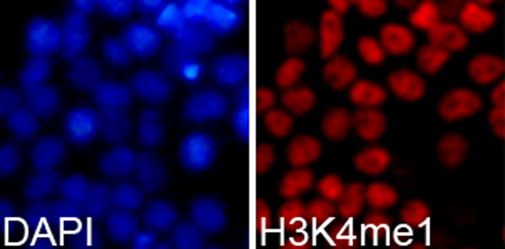
Immunofluorescence analysis of 293T cells using MonoMethyl-Histone H3-K4 antibody (18-618). Blue: DAPI for nuclear staining. |
|
|
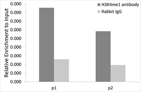
Chromatin immunoprecipitation analysis of extracts of 293T cells, using MonoMethyl-Histone H3-K4 antibody (18-618) and rabbit IgG. P1 and P2 were located on promoter (GAPDH). The amount of immunoprecipitated DNA was checked by quantitative PCR. Histogram was constructed by the ratios of the immunoprecipitated DNA to the input. |
Produktgarantie und fachkundiger Support
