
Symmetric DiMethyl-Histone H3-R26 pAb, Unconjugated, Rabbit, Polyclonal
Artikelnummer:
PRS-18-965
- Bilder (9)
| Artikelname: | Symmetric DiMethyl-Histone H3-R26 pAb, Unconjugated, Rabbit, Polyclonal |
| Artikelnummer: | PRS-18-965 |
| Hersteller Artikelnummer: | 18-965 |
| Alternativnummer: | PRS-18-965-100 |
| Hersteller: | ProSci |
| Wirt: | Rabbit |
| Kategorie: | Antikörper |
| Applikation: | IF, WB |
| Spezies Reaktivität: | Mouse, Rat |
| Immunogen: | A synthetic methylated peptide corresponding to residues surrounding Arg26 of human histone H3 |
| Konjugation: | Unconjugated |
| Alternative Synonym: | H3/b, H3FB, ERBB, ERBB1, HER1, H3/a, H3/c, H3/d, H3/f, H3/h |
| Application Verdünnung: | Optimal dilutions for each application to be determined by the researcher. |
| Anwendungsbeschreibung: | WB: 1:500 - 1:2000IF: 1:50 - 1:200 |
|
|
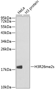
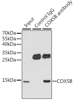
Western blot analysis of extracts of various cell lines, using Symmetric DiMethyl-Histone H3-R26 antibody (18-965). Secondary antibody: HRP Goat Anti-Rabbit IgG (H+L) at 1:10000 dilution. Lysates/proteins: 25ug per lane. Blocking buffer: 3% nonfat dry milk in TBST. |
|
|
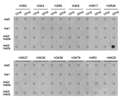
Dot-blot analysis of all sorts of methylation peptides using Symmetric DiMethyl-Histone H3-R26 antibody (18-965). |
|
|
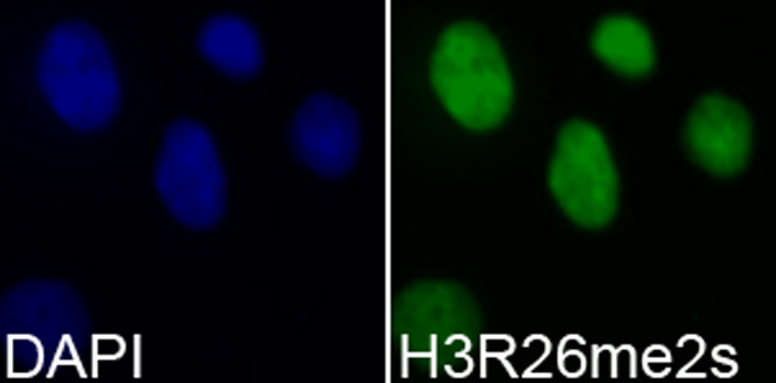
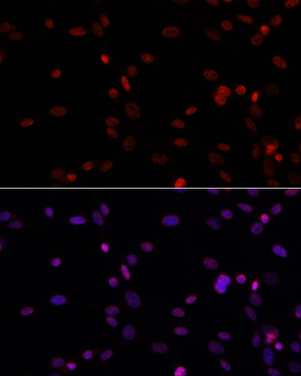
Immunofluorescence analysis of 293T cells using Symmetric DiMethyl-Histone H3-R26 antibody (18-965). Blue: DAPI for nuclear staining. |
|
|
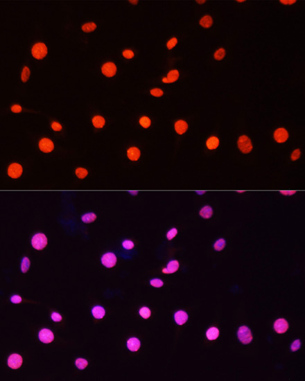
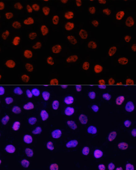
Immunofluorescence analysis of C6 cells using Symmetric DiMethyl-Histone H3-R26 antibody (18-965) at dilution of 1:100. Blue: DAPI for nuclear staining. |
|
|
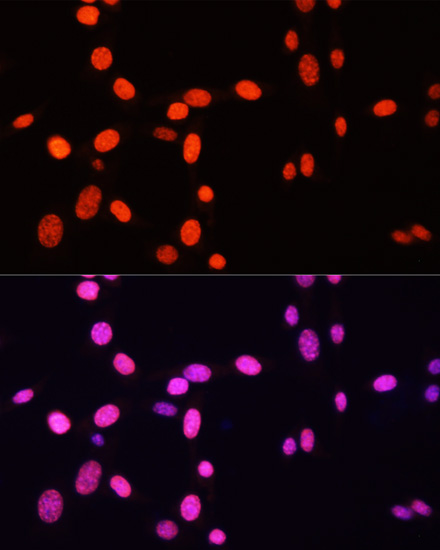
Immunofluorescence analysis of HeLa cells using Symmetric DiMethyl-Histone H3-R26 antibody (18-965) at dilution of 1:100. Blue: DAPI for nuclear staining. |
|
|
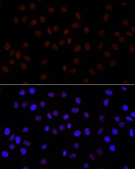
Immunofluorescence analysis of NIH/3T3 cells using Symmetric DiMethyl-Histone H3-R26 antibody (18-965) at dilution of 1:100. Blue: DAPI for nuclear staining. |
|
|
Immunofluorescence analysis of C6 cells using Symmetric DiMethyl-Histone H3-R26 antibody (18-965) at dilution of 1:100. Blue: DAPI for nuclear staining. |
|
|
Immunofluorescence analysis of HeLa cells using Symmetric DiMethyl-Histone H3-R26 antibody (18-965) at dilution of 1:100. Blue: DAPI for nuclear staining. |
|
|
Immunofluorescence analysis of NIH/3T3 cells using Symmetric DiMethyl-Histone H3-R26 antibody (18-965) at dilution of 1:100. Blue: DAPI for nuclear staining. |
Produktgarantie und fachkundiger Support
