
Caspase-9 Antibody, Unconjugated, Rabbit, Polyclonal
Artikelnummer:
PRS-2073
- Bilder (6)

| Artikelname: | Caspase-9 Antibody, Unconjugated, Rabbit, Polyclonal |
| Artikelnummer: | PRS-2073 |
| Hersteller Artikelnummer: | 2073 |
| Alternativnummer: | PRS-2073-0.02,PRS-2073-0.1 |
| Hersteller: | ProSci |
| Wirt: | Rabbit |
| Kategorie: | Antikörper |
| Applikation: | ELISA, ICC, IF, IP, WB |
| Spezies Reaktivität: | Human, Mouse |
| Immunogen: | Anti-Caspase 9 antibody (2073) was raised against a peptide corresponding to 20 amino acids near the center of human Caspase 9. The immunogen is located within amino acids 290-340 of Caspase 9. |
| Konjugation: | Unconjugated |
| Alternative Synonym: | Caspase-9 Antibody: MCH6, APAF3, APAF-3, PPP1R56, ICE-LAP6, MCH6, Caspase-9, Apoptotic protease Mch-6, CASP-9 |
| Application Verdünnung: | Optimal dilutions for each application to be determined by the researcher. |
| Anwendungsbeschreibung: | WB: 1 µg/mL, ICC: 2- 5 µg/mL, IF: 5-20 µg/mL.Antibody validated: Western Blot in human and mouse samples, Immunocytochemistry, Immunofluorescence in human samples. All other applications and species not yet tested. |
|
|
Figure 6 Immunocytochemistry Validation of Caspase 9 in HeLa CellsImmunocytochemical analysis of HeLa cells using anti-Caspase 9 antibody (2073) at 5 &956,g/ml. Cells was fixed with formaldehyde and blocked with 10% serum for 1 h at RT, antigen retrieval was by heat mediation with a citrate buffer (pH6). Samples were incubated with primary antibody overnight at 4&730,C. A goat anti-rabbit IgG H&L (HRP) at 1/250 was used as secondary. Counter stained with Hematoxylin. |
|
|
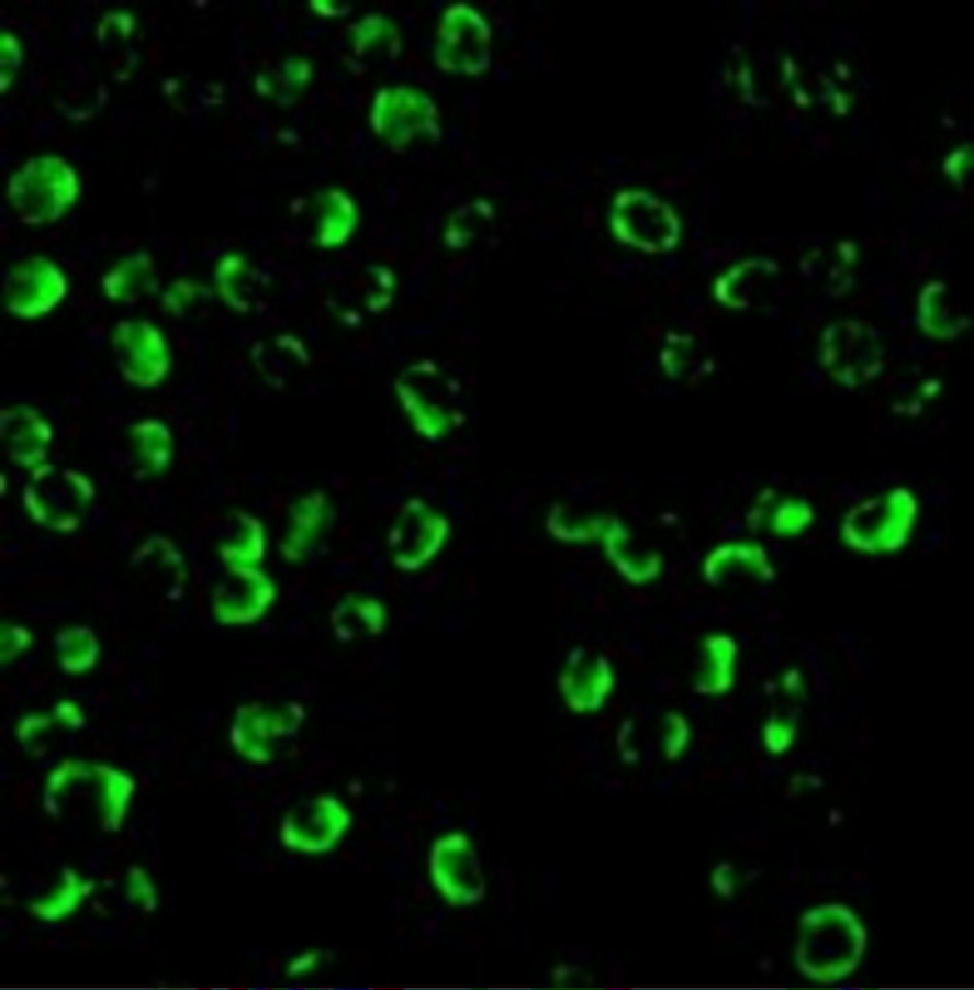
Figure 4 Immunofluorescence Validation of Caspase 9 in K562 CellsImmunofluorescent analysis of 4% paraformaldehyde-fixed K562 cells labeling Caspase 9 with 2073 at 20 &956,g/mL, followed by goat anti-rabbit IgG secondary antibody at 1/500 dilution (green). |
|
|
Figure 5 Immunofluorescence Validation of Caspase 9 in HeLa CellsImmunofluorescent analysis of 4% paraformaldehyde-fixed HeLa cells labeling Caspase 9 with 2073 at 5 &956,g/mL, followed by goat anti-rabbit IgG secondary antibody at 1/500 dilution (green) and DAPI staining (blue). |
|
|
Figure 1 Independent Antibody Validation (IAV) via Protein Expression Profile in Human Cell LinesLoading: 15 &956,g of lysates per lane.Antibodies: Caspase 9, 2071 (1 &956,g/mL), Caspase 9, 2073 (1 &956,g/mL) and beta-actin (1.5 &956,g/mL), 1h incubation at RT in 5% NFDM/TBST.Secondary: Goat anti-rabbit IgG HRP conjugate at 1:10000 dilution. |
|
|
Figure 2 Western Blot Validation in Human Cell LinesLoading: 15 &956,g of lysates per lane.Antibodies: : Caspase 9, 2073 (1 &956,g/mL)), 1h incubation at RT in 5% NFDM/TBST.Secondary: Goat anti-rabbit IgG HRP conjugate at 1:10000 dilution. |
|
|
Figure 3 Western Blot Validation in Mouse Cell LineLoading: 15 &956,g of 3T3/NIH cell lysate.Antibodies: : Caspase 9, 2073 (1 &956,g/mL)), 1h incubation at RT in 5% NFDM/TBST.Secondary: Goat anti-rabbit IgG HRP conjugate at 1:10000 dilution. |
Produktgarantie und fachkundiger Support
