
ACE2 Antibody, Unconjugated, Rabbit, Polyclonal
Artikelnummer:
PRS-3229
- Bilder (10)
| Artikelname: | ACE2 Antibody, Unconjugated, Rabbit, Polyclonal |
| Artikelnummer: | PRS-3229 |
| Hersteller Artikelnummer: | 3229 |
| Alternativnummer: | PRS-3229-0.02,PRS-3229-0.1 |
| Hersteller: | ProSci |
| Wirt: | Rabbit |
| Kategorie: | Antikörper |
| Applikation: | ELISA, IF, IHC-P, WB |
| Spezies Reaktivität: | Human, Mouse, Rat |
| Immunogen: | ACE2 antibody was raised against a synthetic peptide corresponding to amino acids near the center of human ACE2.The immunogen is located within amino acids 150 - 200 of ACE2. |
| Konjugation: | Unconjugated |
| Alternative Synonym: | ACE2 Antibody: ACEH, Angiotensin-converting enzyme 2, ACE-related carboxypeptidase, ACEH, SARS-CoV receptor, SARS-CoV-2 receptor |
| Application Verdünnung: | Optimal dilutions for each application to be determined by the researcher. |
| Anwendungsbeschreibung: | WB: 4 µg/mL, IHC: 2 µg/mL, IF: 20 µg/mL.Antibody validated: Western Blot in human, mouse and rat samples, Immunohistochemistry in human, mouse and rat samples, Immunofluorescence in human, mouse, and rat samples. All other applications and species not yet tested. |
|
|
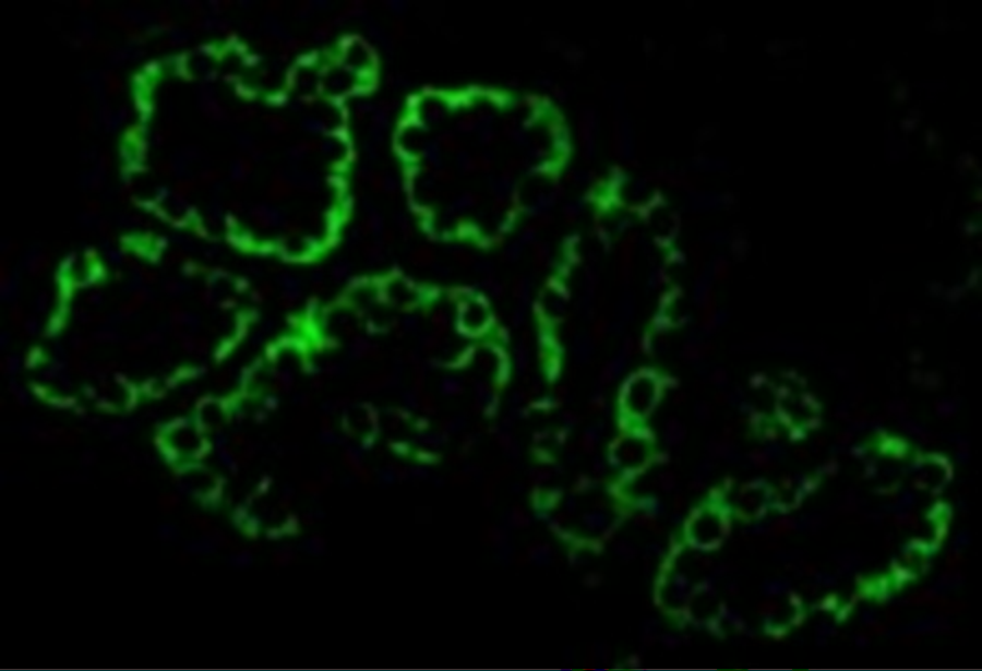
Figure 5 Immunofluorescence Validation of ACE2 in Human Kidney CellsImmunofluorescent analysis of 4% paraformaldehyde-fixed human kidney cells labeling ACE2 with 3229 at 20 &956,g/mL, followed by goat anti-rabbit IgG secondary antibody at 1/500 dilution (green). |
|
|
Figure 6 Immunofluorescence Validation of ACE2 in Human Testis TissueImmunofluorescent analysis of 4% paraformaldehyde-fixed human testis tissue labeling ACE-2 with 3229 at 20 &956,g/mL, followed by goat anti-rabbit IgG secondary antibody at 1/500 dilution (green) and DAPI staining (blue). |
|
|
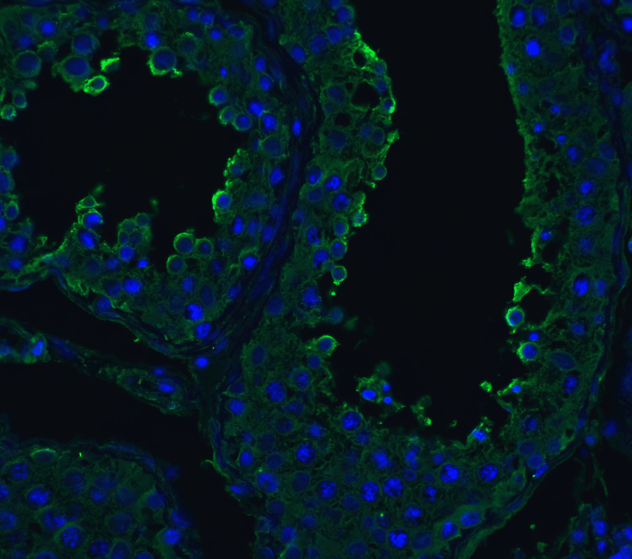
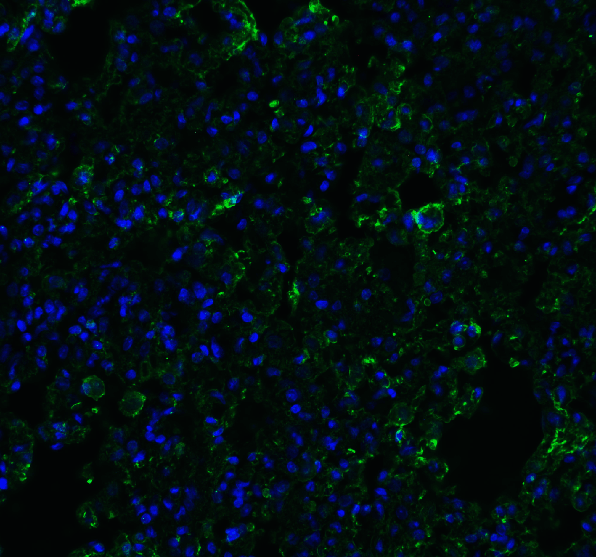
Figure 7 Immunofluorescence Validation of ACE2 in Human Lung TissueImmunofluorescent analysis of 4% paraformaldehyde-fixed human lung tissue labeling ACE-2 with 3229 at 20 &956,g/mL, followed by goat anti-rabbit IgG secondary antibody at 1/500 dilution (green) and DAPI staining (blue). |
|
|
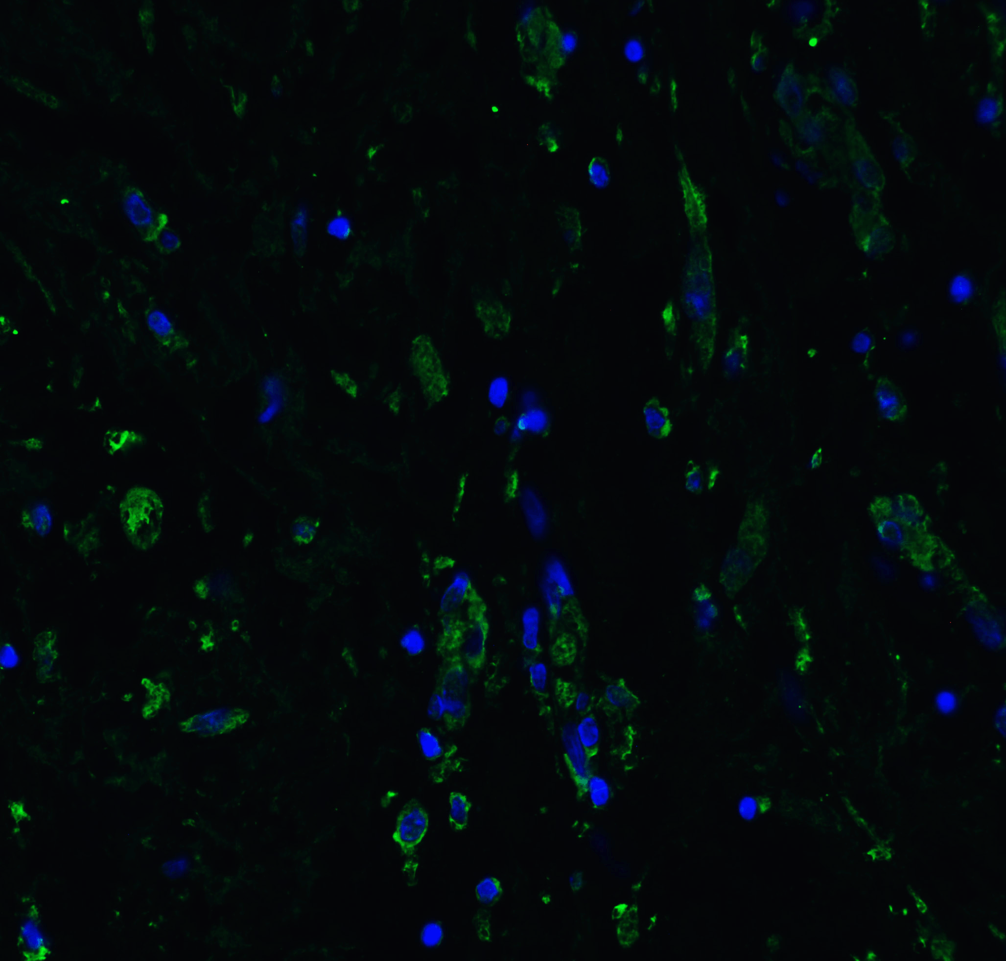
Figure 8 Immunofluorescence Validation of ACE2 in Mouse Lung TissueImmunofluorescent analysis of 4% paraformaldehyde-fixed mouse lung tissue labeling ACE-2 with 3229 at 20 &956,g/mL, followed by goat anti-rabbit IgG secondary antibody at 1/500 dilution (green) and DAPI staining (blue). |
|
|
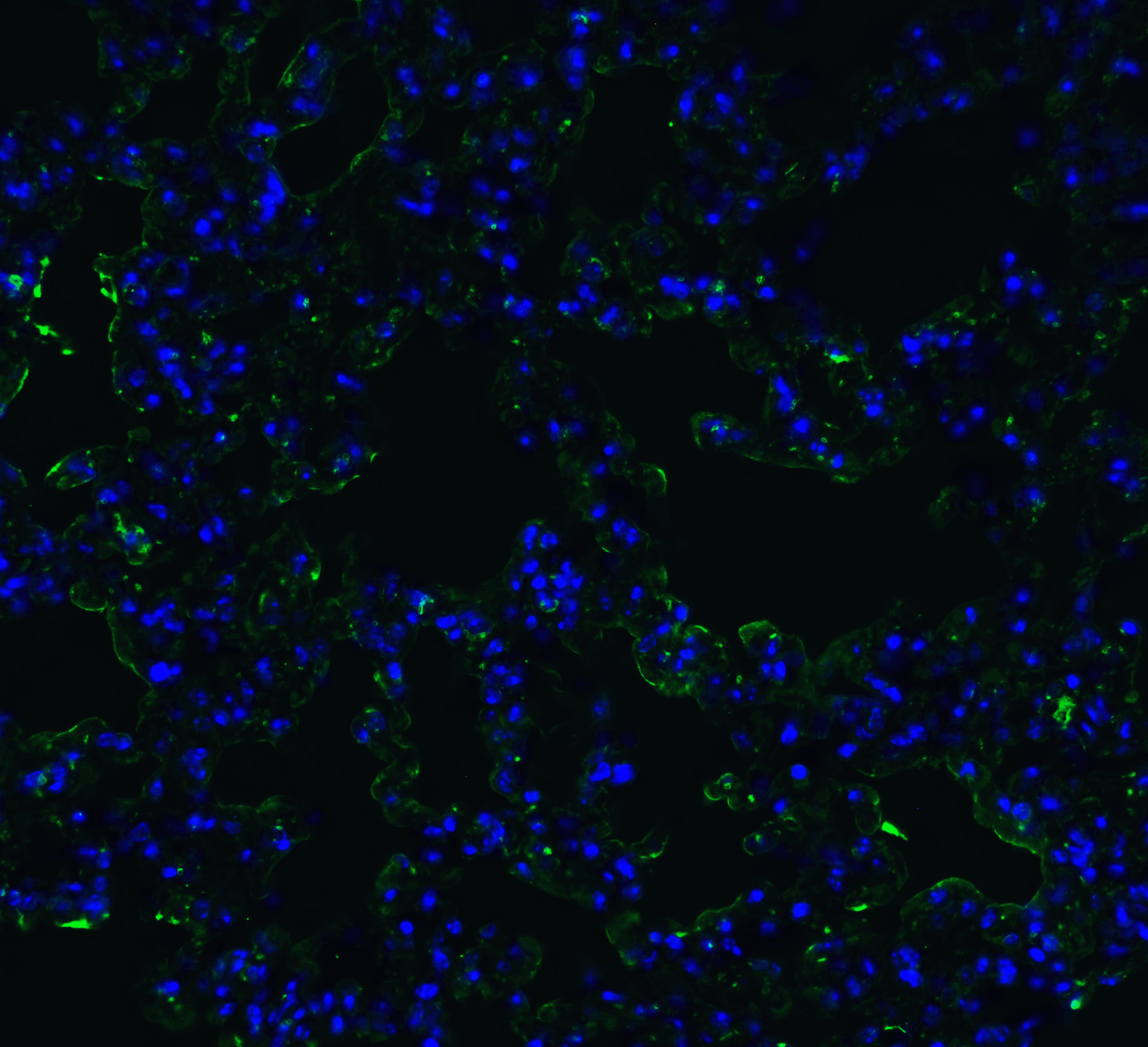
Figure 9 Immunofluorescence Validation of ACE2 in Rat Lung TissueImmunofluorescent analysis of 4% paraformaldehyde-fixed rat lung tissue labeling ACE-2 with 3229 at 20 &956,g/mL, followed by goat anti-rabbit IgG secondary antibody at 1/500 dilution (green) and DAPI staining (blue). |
|
|
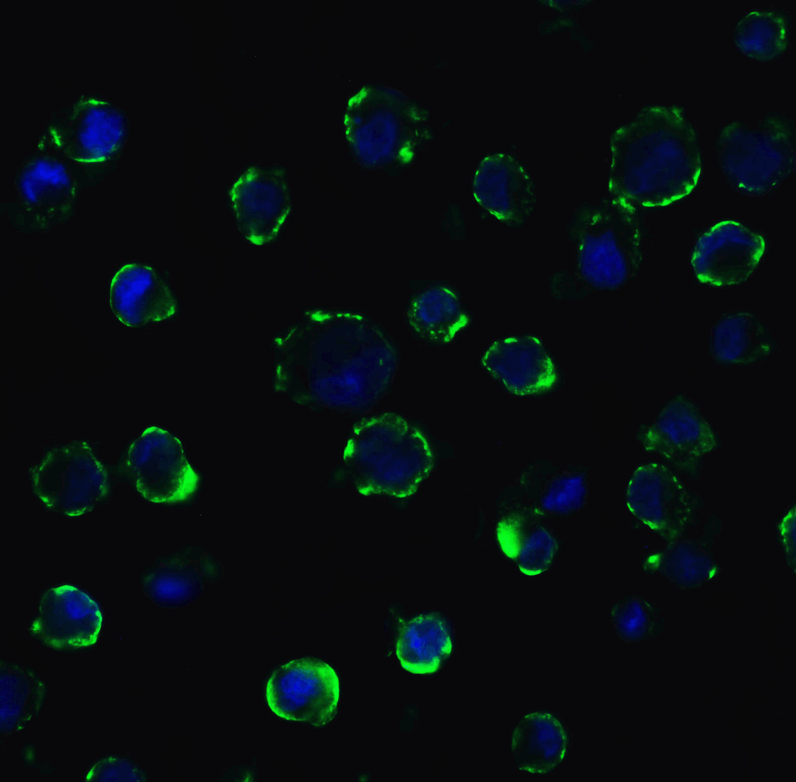
Figure 10 Immunofluorescence Validation of ACE2 In Caco2 Cells Immunofluorescent analysis of 4% paraformaldehyde-fixed Caco2 cells labeling ACE2 with 3229 at 5 &956,g/mL, followed by goat anti-rabbit IgG secondary antibody at 1/500 dilution (green) and DAPI staining (blue). Image showing membrane staining on Caco2 cells. |
|
|
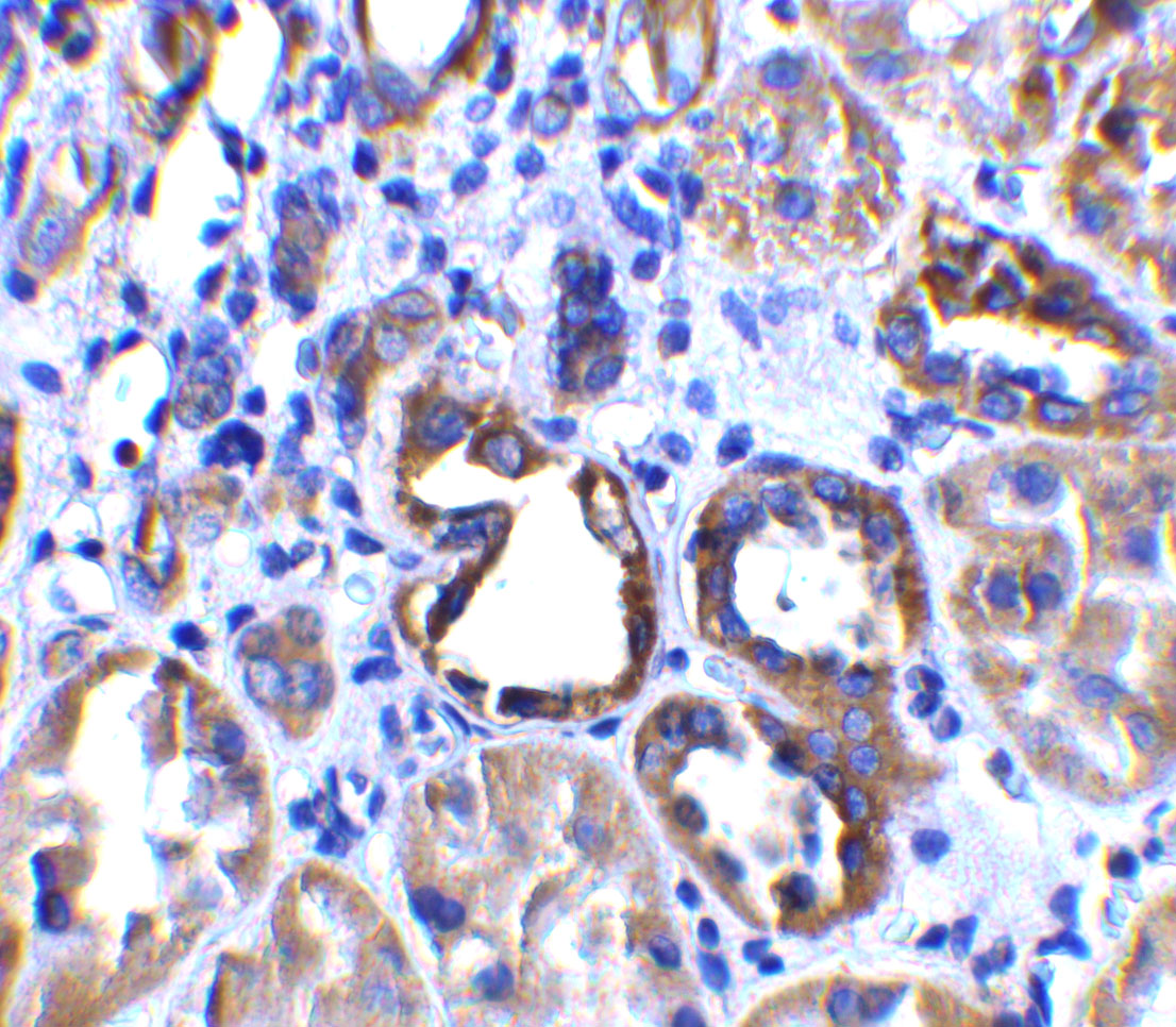
Figure 4 Immunohistochemistry Validation of ACE2 in Human Kidney Tissue Immunohistochemical analysis of paraffin-embedded human kidney tissue using anti-ACE2 antibody (3229) at 2 &956,g/ml. Tissue was fixed with formaldehyde and blocked with 10% serum for 1 h at RT, antigen retrieval was by heat mediation with a citrate buffer (pH6). Samples were incubated with primary antibody overnight at 4 &730,C. A goat anti-rabbit IgG H&L (HRP) at 1/250 was used as secondary. Counter stained with Hematoxylin. |
|
|
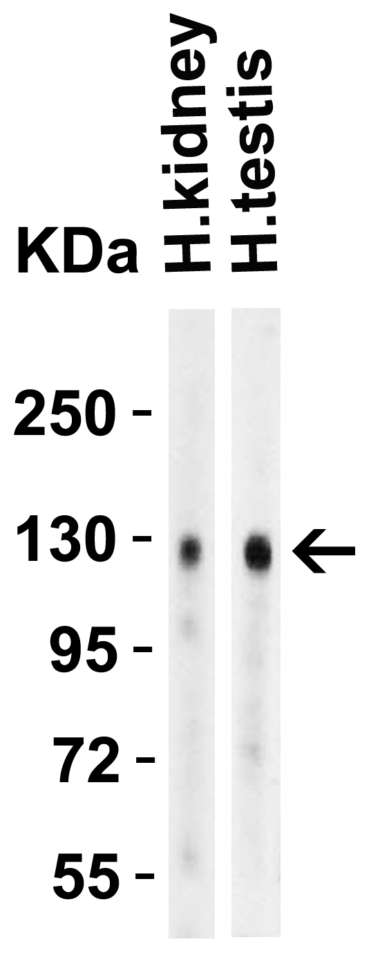
Figure 1 Western Blot Validation in Human TissuesLoading: 15 µg of lysates per lane.Antibodies: ACE2, 3229 (4 &956,g/mL), 1h incubation at RT in 5% NFDM/TBST.Secondary: Goat anti-rabbit IgG HRP conjugate at 1:10000 dilution. |
|
|
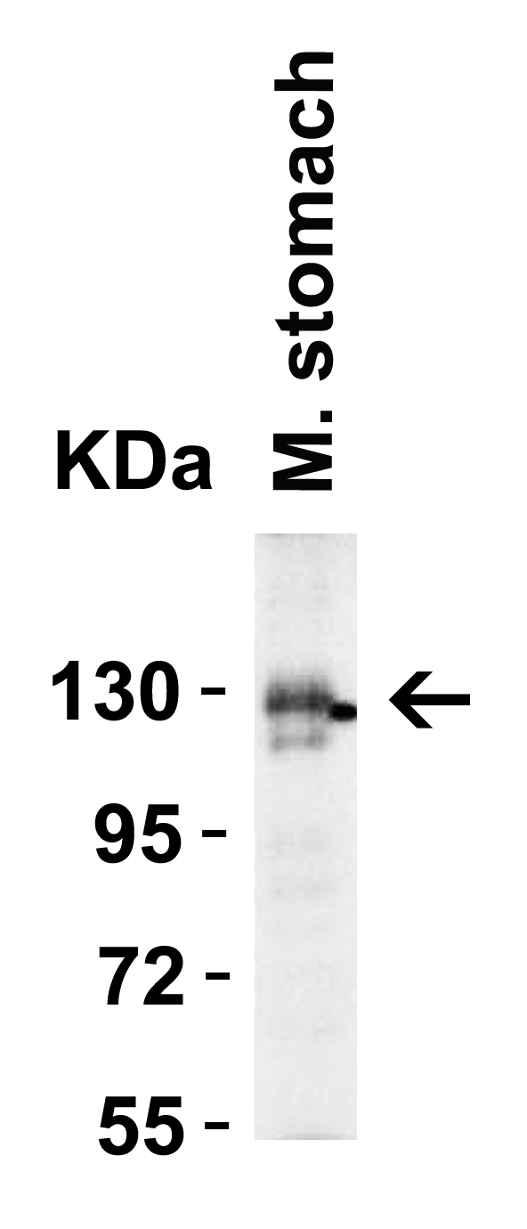
Figure 2 Western Blot Validation in Mouse Stomach TissueLoading: 15 &956,g of lysates per lane.Antibodies: ACE2, 3229 (4 &956,g/mL), 1h incubation at RT in 5% NFDM/TBST.Secondary: Goat anti-rabbit IgG HRP conjugate at 1:10000 dilution. |
|
|
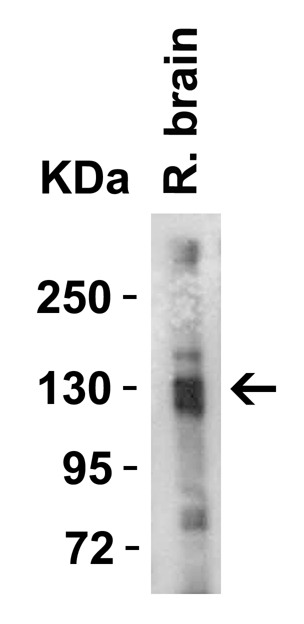
Figure 3 Western Blot Validation in Rat Brain TissueLoading: 15 &956,g of lysates per lane.Antibodies: ACE2, 3229 (4 &956,g/mL), 1h incubation at RT in 5% NFDM/TBST.Secondary: Goat anti-rabbit IgG HRP conjugate at 1:10000 dilution. |
Produktgarantie und fachkundiger Support
