
SARS-CoV-2 (COVID-19) Spike RBD Antibody, Unconjugated, Rabbit, Polyclonal
Artikelnummer:
PRS-9087
- Bilder (11)
| Artikelname: | SARS-CoV-2 (COVID-19) Spike RBD Antibody, Unconjugated, Rabbit, Polyclonal |
| Artikelnummer: | PRS-9087 |
| Hersteller Artikelnummer: | 9087 |
| Alternativnummer: | PRS-9087-0.02,PRS-9087-0.1 |
| Hersteller: | ProSci |
| Wirt: | Rabbit |
| Kategorie: | Antikörper |
| Applikation: | ELISA, IF, IHC, WB |
| Spezies Reaktivität: | Virus |
| Immunogen: | Anti-SARS-CoV-2 (COVID-19) Spike RBD antibody (9087) was raised against a peptide corresponding to 19 amino acids near the carboxy terminus of SARS-CoV-2 (COVID-19) Spike glycoprotein RBD. The immunogen is located within the last 50 amino acids of SARS-CoV-2 (COVID-19) Spike protein RBD. |
| Konjugation: | Unconjugated |
| Alternative Synonym: | SARS-CoV-2 (COVID-19) Spike RBD Antibody: Severe acute respiratory syndrome coronavirus 2 (SARS-CoV-2), Spike protein, Surface Glycoprotein, covid-19, sars-cov-2 |
| Application Verdünnung: | Optimal dilutions for each application to be determined by the researcher. |
| Anwendungsbeschreibung: | WB: 2-5 µg/mL, IF: 20 µg/mL, IHC: 0.5 µg/mL.Antibody validated: Western Blot in human samples, Immunohistochemistry and Immunofluorescence in human samples. SARS-CoV-2 (COVID-19) Spike RBD Antibody can be used for the detection of SARS-CoV-2 (COVID-19) Spike protein in ELISA and WB. It will detect 4 ng of free peptide at 1 µg/mL. All other applications and species not yet tested. |
|
|
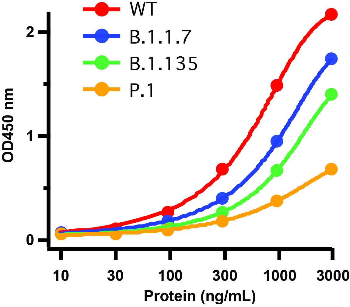
Figure 1 Detection of SARS-CoV-2 Variant Proteins with Spike RBD Antibodies by Direct ELISA Coating Antigen: SARS-CoV-2 full length spike proteins, including WT, UK variant (B.1.1.7), SA variant (B.1.135) and Brazil (P.1). Dilution: 10-3000 ng/mL. Incubate at 4 &730,C overnight.Detection Antibodies: SARS-CoV-2 Spike RBD Antibody, 9087, 2 &956,g/mL, incubate at RT for 1 hr.Secondary Antibodies: Goat anti-rabbit HRP at 1:20,000, incubate at RT for 1 hr.Immunogen region of antibody (9087) includes site 501N that was mutated in all three variants. 9087 has low binding affinity for all three variants as compared to WT. |
|
|
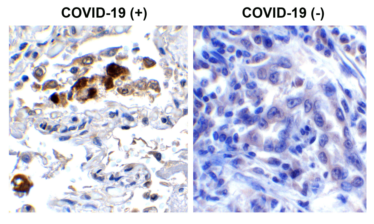
Figure 2 Immunohistochemistry Validation of SARS-CoV-2 (COVID-19) Spike RBD in COVID-19 Patient Lung Immunohistochemical analysis of paraffin-embedded COVID-19 patient lung tissue using anti- SARS-CoV-2 (COVID-19) Spike RBD antibody (9087, 0.5 &956,g/mL). Tissue was fixed with formaldehyde and blocked with 10% serum for 1 h at RT, antigen retrieval was by heat mediation with a citrate buffer (pH6). Samples were incubated with primary antibody overnight at 4&730,C. A goat anti-rabbit IgG H&L (HRP) at 1/250 was used as secondary. Counter stained with Hematoxylin. Strong signal of SARS-COV-2 Spike RBD protein was observed in macrophage of COVID-19 patient lung, but not in non-COVID-19 patient lung. |
|
|
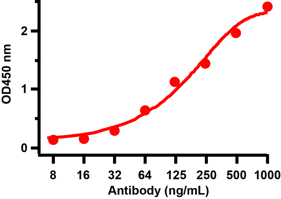
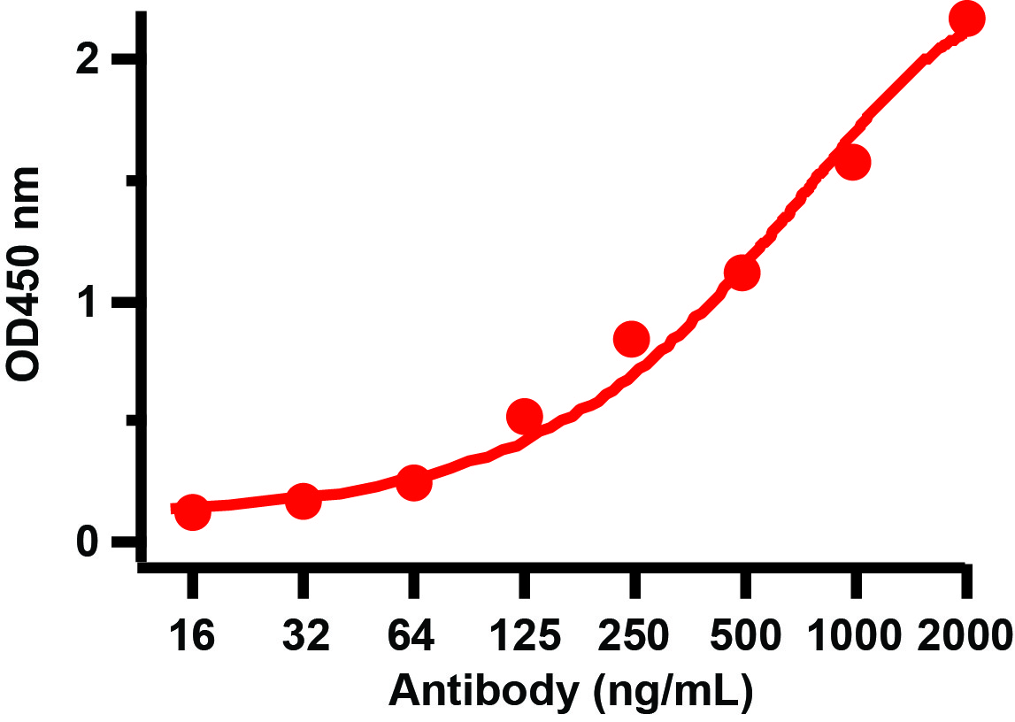
Figure 4 ELISA Validation with SARS-CoV-2 (COVID-19) Spike RBD Recombinant ProteinAntibodies: SARS-CoV-2 (COVID-19) Spike RBD antibody, 9087 (1 &956,g/mL). A direct ELISA was performed using SARS-CoV-2 (COVID-19) Spike RBD recombinant protein (10-303) as coating antigen and the anti-SARS-CoV-2 (COVID-19) Spike RBD antibody as the capture |
|
|
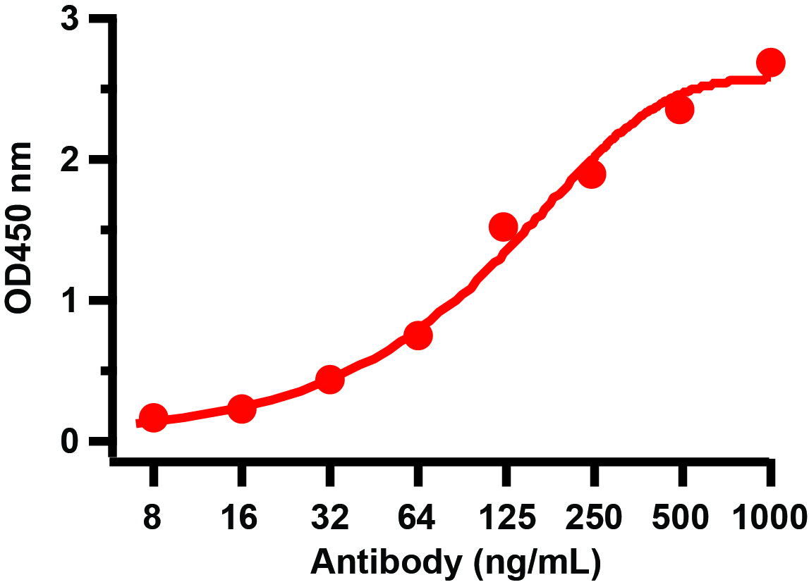
Figure 8 ELISA Validation with SARS-CoV-2 (COVID-19) Spike RBD+SD1 Recombinant ProteinAntibodies: SARS-CoV-2 (COVID-19) Spike RBD antibody, 9087 (1 &956,g/mL). A direct ELISA was performed using SARS-CoV-2 (COVID-19) Spike RBD+SD1 recombinant protein (10-304) as coating antigen and the anti-SARS-CoV-2 (COVID-19) Spike RBD antibody as the capture antibody. Secondary: Goat anti-rabbit IgG HRP conjugate at 1:20000 dilution. Detection range is from 8 ng/mL to 1000ng/mL. |
|
|
|
|
|
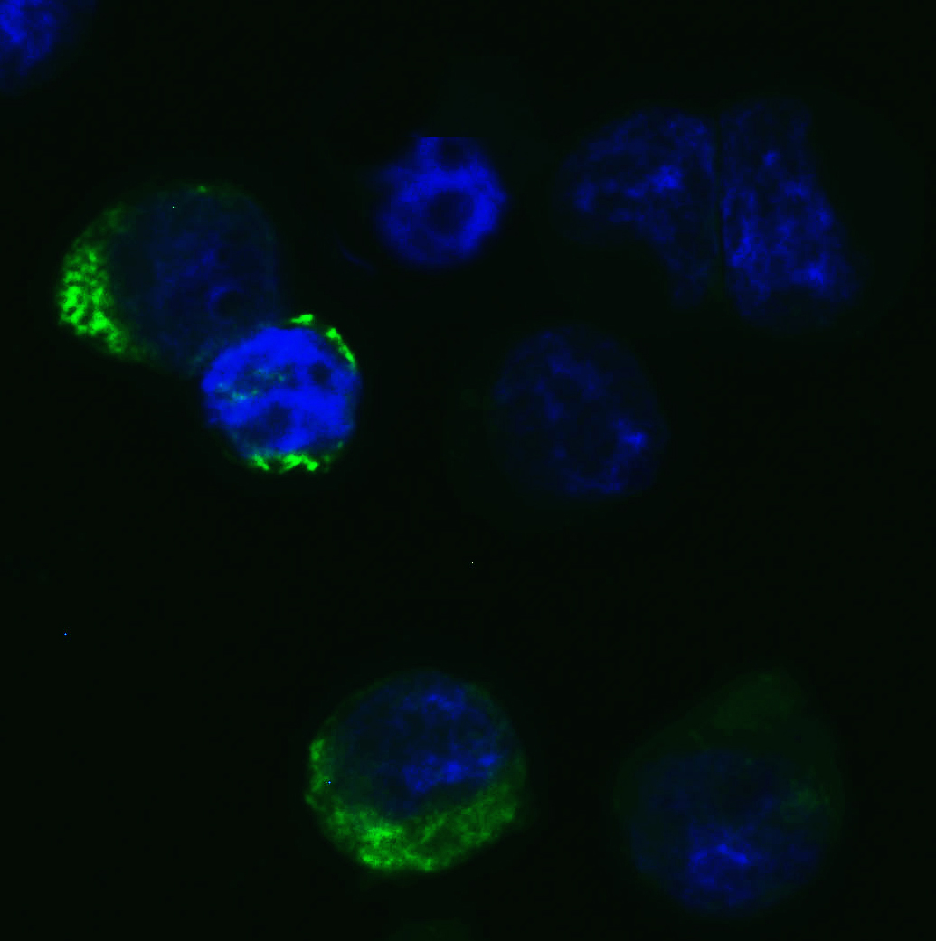
Figure 5 Immunofluorescence Validation of SARS-CoV-2 (COVID-19) Spike RBD in 293 Transfected CellsImmunofluorescent analysis of 4% paraformaldehyde-fixed Spike transfected 293 cells labeling SARS-CoV-2 (COVID-19) Spike RBD with 9087 at 20 ug/mL, followed by goat anti-rabbit IgG secondary antibody at 1/500 dilution (green) and DAPI staining (blue). |
|
|
Figure 3 Western Blot Validation with SARS-CoV-2 (COVID-19) Spike RBD Recombinant Protein Loading: 30 ng per lane of SARS-CoV-2 (COVID-19) Spike RBD recombinant protein, 10-303. Antibodies: SARS-CoV-2 (COVID-19) Spike RBD, 9087, 1h incubation at RT in 5% NFDM/TBST. Secondary: Goat anti-rabbit IgG HRP conjugate at 1:10000 dilution. Lane 1: 1 µg/mL and Lane 2: 2 µg/mL. |
|
|
Figure 6 Overexpression Validation in Spike Transfected 293 Cells Loading: 10 &956,g per lane of 293 cell lysate. Antibodies: SARS-CoV-2 (COVID-19) Spike RBD, 9087 (4 &956,g/mL), 1h incubation at RT in 5% NFDM/TBST. Secondary: Goat anti-rabbit IgG HRP conjugate at 1:10000 dilution. Lane 1: WT 293 cells and Lane 2: SARS-CoV-2 Spike overexpressed 293 cells |
|
|
Figure 7 Western Blot Validation with SARS-CoV-2 (COVID-19) Spike RBD+SD1 Recombinant ProteinLoading: 30 ng per lane of SARS-CoV-2 (COVID-19) Spike RBD+SD1 recombinant protein, 10-304. Antibodies: SARS-CoV-2 (COVID-19) Spike RBD, 9087, 1h incubation at RT in 5% NFDM/TBST. Secondary: Goat anti-rabbit IgG HRP conjugate at 1:10000 dilution. Lane 1: 1 µg/mL and Lane 2: 2 µg/mL. |
|
|
Figure 9 Western Blot Validation with SARS-CoV-2 (COVID-19) Spike RBD+SD1+SD2 Recombinant ProteinLoading: 30 ng per lane of SARS-CoV-2 (COVID-19) Spike RBD+SD1+SD2 recomb |
|
|
Produktgarantie und fachkundiger Support
