0.02 M Potassium Phosphate, 0.15 M Sodium Chloride, pH 7.2
Quelle:
Human
Formulierung:
Liquid (sterile filtered)
Application Verdünnung:
ELISA: 1:5000 - 1:50,000, WB: 1:1000
Anwendungsbeschreibung:
HUMAN IgG F(c) Fragment has been tested by SDS-Page and dot blot and can be utilized as a control or standard reagent in SDS, Western Blotting, and ELISA experiments.
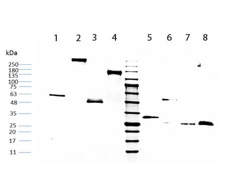
SDS-PAGE of Human IgG Fc (009-0103). Lane 1: Non-reduced Human IgG Fc. Lane 2: Non-reduced Human IgG Whole Molecule (009-0102). Lane 3: Non-reduced Human IgG Fab Fragment (009-0105). Lane 4: Non-reduced Human IgG F(ab)2 (009-0104). Middle Lane: 5µL OPAL Pre-stained Marker MB-210-0500. Lane 5: Reduced Human IgG Fc. Lane 6: Reduced Human IgG Whole Molecule (009-0102). Lane 7: Reduced Human IgG Fab Fragment (009-0105). Lane 8: Reduced Human IgG F(ab)2 (009-0104). Load: 1µg per lane. Predicted/Observed size: Non-reduced at 50kDa, reduced at 25kDa/Non-reduced at 55-60kDa, reduced at 30-33kDa.
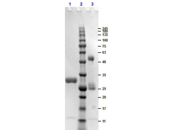
SDS-PAGE results of Human IgG F(c) Fragment. Lane 1: reduced Human IgG F(c) Fragment. Lane 2: Opal Prestained Molecular Weight Ladder (p/n MB-210-0500). Lane 3: non-reduced Human IgG F(c) Fragment. Load: 1µg. 4-20% Lonza SDS-PAGE, Coomassie Stained, BioRad ChemiDoc Imaged.
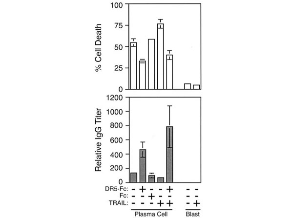
TRAIL-mediated apoptosis of human plasma cells.Top,Control CESS cells (blast) or IL-6-differentiated plasma cells were cultured for 24 h in the presence or absence of 1.0 µg/ml TRAIL, DR5-Fc, or the Fc portion of human IgG. Cell viability was determined by trypan blue exclusion, and the percent of plasma cell death was calculated by dividing the number of dead plasma cells by the total number of plasma cells in the culture.Bottom,ELISA of IgG secretion in the corresponding culture medium. Data represent the relative IgG titer required to obtain a single OD value that lies within the linear range. Data represent three independent cell populations from one experiment. Three independent experiments were performed with similar results. Fig 3. PMID: 12421926.
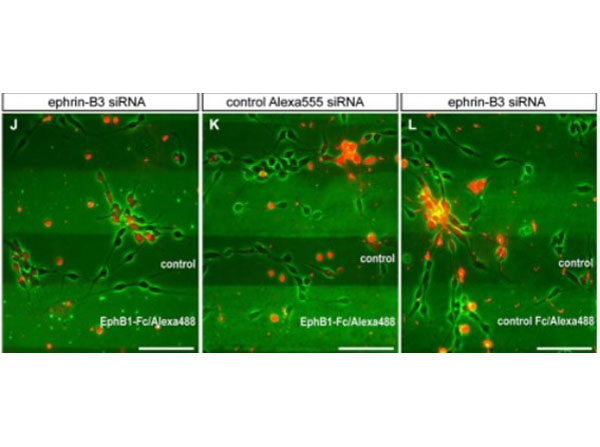
EphB1 acts repulsive on neurons of the superficial migratory stream via reverse signaling with stripe assay.(J)After down regulation of ephrin-B3 ligands by siRNA transfection (red) in MGE-derived neurons the repulsion induced by EphB1-Fc in the stripe assay is abolished after 2 DIV, while non-transfected interneurons still avoid the EphB1-Fc stripes. (K) Application of Alexa555 labeled control siRNA has no effect as most of the cells still avoid the EphB1-Fc containing stripes. (L) Addition of ephrin-B3 siRNA does not affect MGE-derived neurons growing on Fc/control stripes. Scale bars:(J-L)50 µm. Figure 3. PMID: 25100946.
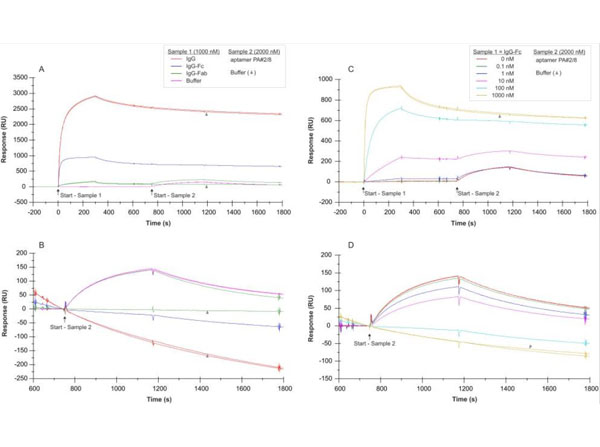
SPR interaction analyses regarding the aptamer binding site in Protein A.Biacore X100 / sensor chip CAP / ligand: biotinylated Protein A with immobilization level of ~560 RU / two-step analyte binding without regeneration in between, (A-B) analyte 1 = sample 1: human IgG, IgG-Fc fragm
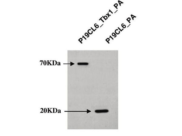
Western blot analysis of P19CL6_Tbx1-PA cells and P19CL6_PA control cells. Proteins were separated on 10% SDS-PAGE gel and immunoblotted with human IgG F(c). Figure 1. PMID: 19178302.
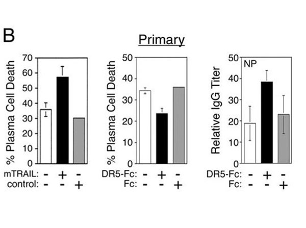
TRAIL induces apoptosis of primary plasma cells generated in a T-dependent immune response.(B)The in vivo-generated, syndecan-1-positive plasma cells were cultured alone (□) or with TRAIL-expressing 2PK3 cells (mTRAIL, ▪) or the parental 2PK3 (control, ▦) cells at a 10:1 ratio for 5 h before determination of cell death by trypan blue staining (left panel). In vivo-generated plasma cells were plated onto CD40L-expressing L cells at a 10:1 ratio in the absence (□) or presence (▪) of DR5-Fc or Fc (▦) (1 µg/ml). Cell viability was determined by trypan blue exclusion after a 5-h incubation (middle panel), and the relative amount of NP-specific IgG secreted into the medium for a total of 20 h was determined by ELISA (right panel).For each of the conditions, the values represent independent analysis of cells isolated from three mice. Fig 4. PMID: 12421926.
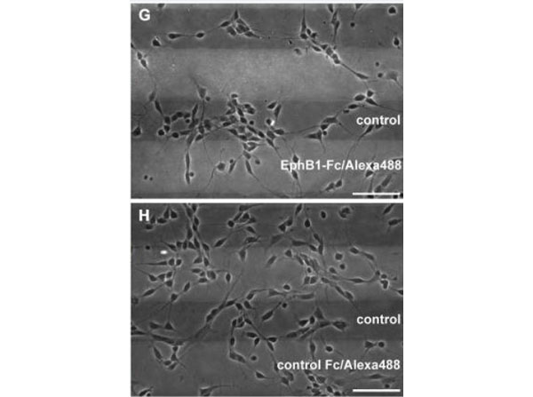
EphB1 acts repulsive on neurons of the superficial migratory stream via reverse signaling with stripe assay. (G) Dissociated neurons from the MGE clearly avoid EphB1-containing lanes in the stripe assay after 2 DIV. (H) On alternating stripes of labeled and unlabeled control protein the cells show no preferential growth. Scale bars:(G-H)50 µm. Figure 3. PMID: 25100946.
* Mehrwertsteuer und Versandkosten nicht enthalten. Irrtümer und Preisänderungen vorbehalten