The whole rabbit serum was prepared by repeated immunizations with a synthetic peptide corresponding to amino acids 805-820 of mouse Gli-1. The peptide was synthesized as a multiple antigen peptide (MAP).
Konjugation:
Unconjugated
Alternative Synonym:
rabbit anti-Gli-1 Antibody, rabbit anti-Gli1 Antibody, Zinc finger protein GLI1 antibody, Glioma-associated oncogene antibody, Oncogene GLI antibody
This antibody has been tested for use in ELISA, Immunofluorescence, and western blot. Specific conditions for reactivity should be optimized by the end user. Expect a band approximately 120 kDa in size corresponding to Gli-1 protein by western blotting i
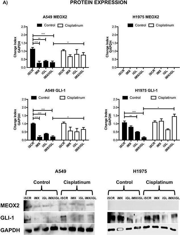
Inducible MEOX2-GLI1 axis expression was involved in cellular migration and cellular proliferation in lung cancer cells(A) A549 and H1975 lung cancer cells demonstrated an inducible GLI-1 protein expression pattern following treatment with 8 µM cisplatinum and reduced GLI-1 inducible expression following the application of specific anti-MEOX2 siRNA and/or anti-MEOX2 siRNA plus anti-GLI1 siRNA in the presence of 8 µM cisplatinum. Western blot statistical analyses, assessed via quantitative densitometry, were performed to determine *p0.05 by one-way ANOVA and Dunnetts test for multiple comparisons to identify significant differences with respect to controls. Students t-test was performed to identify significant differences between control and cisplatinum treatment. Quantification analyses were normalized to scrambled siRNA as a negative control for gene silencing. Images were obtained using a C-DIGIT device (LICOR), and pixel quantification and data analyses were carried out using Image Studio software. Total pixel intensity for each specific protein product was normalized to GAPDH. (B) Cell culture images and graphs showing the quantitative analysis of cellular migration as a percentage (transwell migration assays) indicated significant MEOX2 and GLI-1 protein-dependent functions following the individual and combined application of anti-MEOX2 and anti-GLI1 siRNAs in A549, NH2347 and H1975 lung adenocarcinoma cells, **p0.005 and ***p0.0001 based on one-way ANOVA and Dunnetts multiple comparisons te
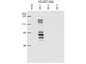
Western blot using Rocklands anti-Gli-1 antibody shows detection of a band at ~150 kDa corresponding to Gli-1 present in HEK293T whole cell lysate transiently transfected with Gli-1 (lane 2). Mock 293T cell lysates and 293T cell lysates transfected with Gli-2 and Gli-3 show no staining (lanes 1, 3 and 4 respectively). Lysates were separated by SDS-PAGE and transferred to nitrocellulose. After blocking, the membrane was probed with Anti-Gli-1 antiserum diluted 1:5,000, followed by HRP conjugated donkey anti-rabbit (Rockland p/n 611-7302). Personal communication, Tom Curran, Childrens Hospital of Philadelphia, Philadelphia, PA.
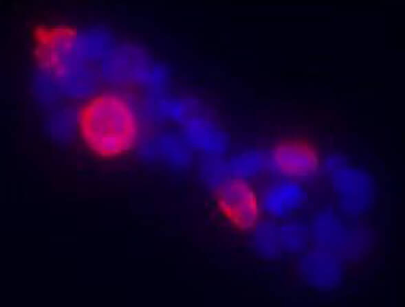
Immunofluorescence of Anti-Gli-1 antibody. HEK293T cells were transiently transfected with Gli-1 (murine). Primary Antibody: Rocklands Anti-Gli-1 antiserum (rabbit) was added 1:400. Secondary Antibody: fluorescent labeled anti-rabbit IgG. Personal communication, Tom Curran, Childrens Hospital of Philadelphia, Philadelphia, PA. Detection of mouse Gli-1 present in transfected 293T cells (red).
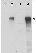
Western blot using Rocklands anti-Gli-1 antibody shows detection of a band at ~150 kDa (arrowhead) corresponding to human Gli-1 present in transfected 293T cell lysates (lanes 2 and 4). Mock 293T cell lysates with vector only show no staining (lanes 1 and 3). Lysates were separated by SDS-PAGE and transferred to nitrocellulose. After blocking the membrane was probed with the primary antibody diluted to 1:8,000 (lanes 1 and 2) or 1:4,000 (lanes 3 and 4). Molecular weight estimation was made by comparison to MW markers. Personal communication, Hiro Kimura, St. Jude Childrens Research Hospital, Memphis, TN.
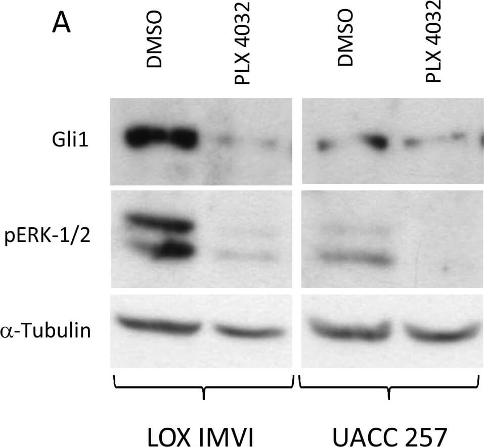
Inhibition of BRAFV600E, expression of GLI1 and SHH-GLI pathway inhibition by NVP-LDE225 in human melanoma cells in vitro. A) LOX IMVI and UACC 257 with PLX-4032 at the dose of 1 µM for 24 hr. subsequently protein lysates prepared and subjected to WB analysis for the expression of GLI1 and phospho-ERK1/2. B) Effect of NVP-LDE225 on PTCH1 promoter. In total, 1 µg of PTCH1 pGL3b-hPTCH1-prom-wt or pGL3b-hPTCH1-prom-mut luciferase construct and reporter were cotransfected into LOX IMVI cells. Cells were subsequently treated with 10 µM of NVP-LDE225 or cyclopamine for 4 hours (time point selected base on kinetic experiments). Fold activation was calculated relative to cells transfected with 3 µg of pB-actin-RL. One representative experiment of 2 is shown. Figure provided by CiteAb. Source: PLoS One, PMID: 23935925.
* Mehrwertsteuer und Versandkosten nicht enthalten. Irrtümer und Preisänderungen vorbehalten