AKT Antibody was produced from whole rabbit serum prepared by repeated immunizations with a synthetic peptide R-P-H-F-P-Q-F-S-Y-S-A-S-G-T-A corresponding to the C-terminus (460-480) of human AKT proteins conjugated to KLH using maleimide. A residue of cysteine was added to the amino terminal end to facilitate coupling. A BLAST analysis was used to suggest reactivity with this protein from rat, mouse, and chicken based on 100% homology for the immunogen sequence.
Anti-AKT Antibody has been tested in Western Blot, Immunohistochemistry (Formalin-fixed paraffin-embedded sections), and Immunofluorescence (paraformaldehyde-fixed primary cardiomyocyte cultures). Expect a band at ~55.7kDa in 3T3 whole cell lysate or oth
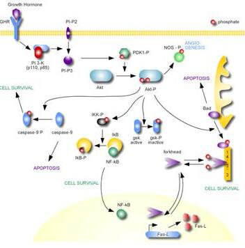
AKT Metabolic Pathway
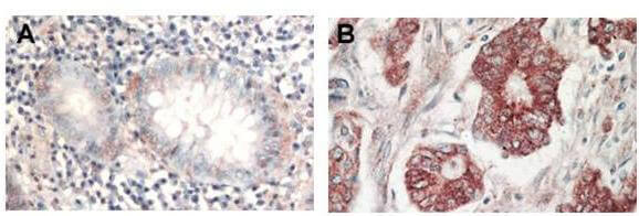
Immunohistochemistry of Rabbit Anti-AKT antibody. Tissue: (A) normal colon tissue, (B) colon tumor tissue. Fixation: formalin fixed paraffin embedded. Antigen retrieval: not required. Primary antibody: AKT antibody at 1:1,000 dilution for 1 h at RT. Secondary antibody: Peroxidase rabbit secondary antibody at 1:10,000 for 45 min at RT. Localization: AKT is nuclear. Staining: AKT as precipitated red signal with hematoxylin purple nuclear counterstain.
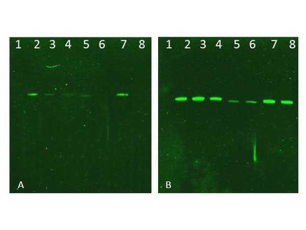
Western Blot of Rabbit AKT Antibodies. Lane 1: NIR MW protein ladder. Lane 2: AKT1, recombinant: 009-001-P21. Lane 3: AKT1, phosphatase-treated: 009-001-I51. Lane 4: AKT1, mutant T308A/S473A: 009-001-P22. Lane 5: AKT2, recombinant: 009-001-P23. Lane 6: AKT2, phosphatase-treated: 009-001-E71. Lane 7: AKT3, recombinant: 009-001-P24. Lane 8: AKT3, phosphatase-treated: 009-001-E75. Load: 50ng per lane. Blot A: 600-401-269 Anti-Akt pT308 used at 1:2270, Blot B: 100-401-401 Anti-Akt used 1:1000.
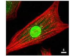
Immunofluorescence Microscopy of Rabbit Anti-AKT Antibody. Tissue: neonatal rat cardiomyocytes. Fixation: 0.5% PFA. Antigen retrieval: not required. Primary antibody: AKT antibody at 1:80 dilution for 1 h at RT. Secondary antibody: Texas-red(TM) conjugated rabbit secondary antibody at 1:10,000 for 45 min at RT. Localization: AKT is nuclear. Staining: Anti-AKT staining appears green. Actin filaments are labeled red using a Texas-red(TM) conjugated phalloidin.
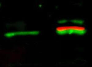
Western Blot of simultaneous detection of unphosphorylated and phosphorylated Rabbit Anti-AKT antibody. Lane 1: unstimulated NIH/3T3 lysates contain inactive unphosphorylated Akt1, green band. Lane 2: PDGF stimulated NIH/3T3 lysate contains both inactive (green band) and activated phosphorylated Akt1 (red band). Load: 35 µg per lane. Primary antibody: rabbit anti-Akt (pan) and mouse anti-Akt pS473 specific antibodies at 1:1000 for overnight at 4C. Secondary antibody: DyLight(TM) 549 conjugated anti-rabbit IgG (green) and DyLight(TM) 649 conjugated anti-mouse IgG (red) secondary antibodies at 1:10,000 for 45 min at RT. Block: 5% BLOTTO overnight at 4C.
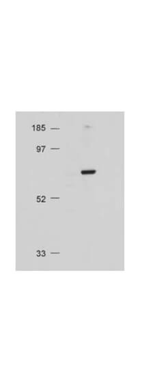
Western Blot of Rabbit Anti-AKT antibody. Lane 1: Molecular Weight. Lane 2: NIH/3T3 whole cell lysate. Load: 20 µg lysate per lane. Primary antibody: Anti-AKT antibody at 1:500 for overnight at 4C. Secondary antibody: HRP conjugated GT-a-Rabbit IgG (611-103-122) at 1:10,000 preceded color development using Pierce Chemicals SuperSignal(TM) substrate. Block: MOPS buffer overnight at 4C. Predicted/Observed size: 56 kDa, 56 kDa for AKT. Other band(s): none.
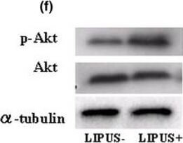
Western blotting analysis. (a) Type-II collagen. (b) Type-IX collagen. (c) Focal adhesion kinase (FAK) and phosphorylated FAK (p-FAK). (d) Paxillin and phosphorylated Paxillin (p-Paxillin). (e) Mitogen-activated protein kinase (MAPK) and phosphorylated MAPK (p-MAPK). There are no evident differences in the expression levels of total MAPK and p-MAPK between the two groups. (f) Akt and phosphorylated Akt (p-Akt). There were no differences found in the intensity the total Akt expression between the two groups, but p-Akt was found at higher levels in the LIPUS group (US+) in comparison with the control group (US-). (g) Cyclin B1 and cyclin D1. (h) Changes of proliferating cell nuclear antigen (PCNA) using MEK1 inhibitor (PD98059) and phosphatidylinositol 3-OH kinase (PI3K) inhibitor (LY294002). Chondrocytes were pretreated with MEK1 inhibitor (PD98059, 250 µM/ml) and PI3K inhibitor (LY294002, 250 µM/ml) for 12 hours and 24 hours followed by stimulation with LIPUS for 20 minutes. Each sample was harvested 2 hours after LIPUS stimulation and the influence of these inhibitors was judged in western blotting analysis of the expression of PCNA. Figure provided by CiteAb. Source: Arthritis Res Ther, PMID: 18616830.
* Mehrwertsteuer und Versandkosten nicht enthalten. Irrtümer und Preisänderungen vorbehalten