This whole rabbit serum was prepared by repeated immunizations with a synthetic peptide corresponding to a N-terminus region near aa 15-45 of mouse angiopoietin 1 protein conjugated to KLH using maleimide. A residue of cysteine was added to the amino terminal end to facilitate coupling.
Anti-Antiopoietin-1 Antiserum has been tested in ELISA, western blot, immunohistochemistry, and immunofluorescence and is suitable for other antibody based assays. A 1:500 dilution is recommended for western blotting. The reaction of this antiserum direc
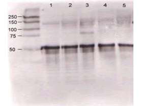
Rabbit anti-Ang-1 was used at a 1:500 dilution to detect mouse Ang-1 by western blot against supernatants of mouse angiopoietin-expressing endothelial cells. Lane 1 - wt endothelial cells. Lane 2 - mouse Ang-1 (clone 1-8) expressing cells. Lane 3 - mouse Ang-1 (clone 1-15) expressing cells. Lane 4 - mouse Ang-2 (clone 2-9) expressing cells. Approximately 20 µg of each lysate was used for 10% SDS-PAGE. Immunoprecipitation preceded the reaction with primary antibody at room temperature for 1 h. After subsequent washing, a 1:5,000 dilution of HRP conjugated Gt-a-Rabbit IgG (611-103-122) preceded color development.
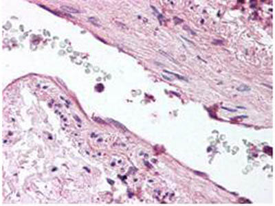
Rocklands anti-ANG1 antibody was diluted 1:500 to detect ANG1 in human lung tissue. Tissue was formalin fixed and paraffin embedded. No pre-treatment of sample was required. The image shows the localization of antibody as the precipitated red signal, with a hematoxylin purple nuclear counter stain.
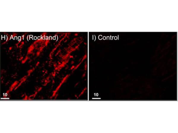
Immunofluorescence and confocal imaging of Rabbit Anti-Angiopoietin 1 Antibody. (H) Perfusion-fixed mouse Left Ventricle. Anti-Angiopoietin 1 conjugated to Alexa Fluor 568 (red), which recognizes only monomers, immunostains CMs and ECs in vivo. I) Rabbit sera (negative control). Scale bars = 10 µm. Dallabrida SM et al. 2008.
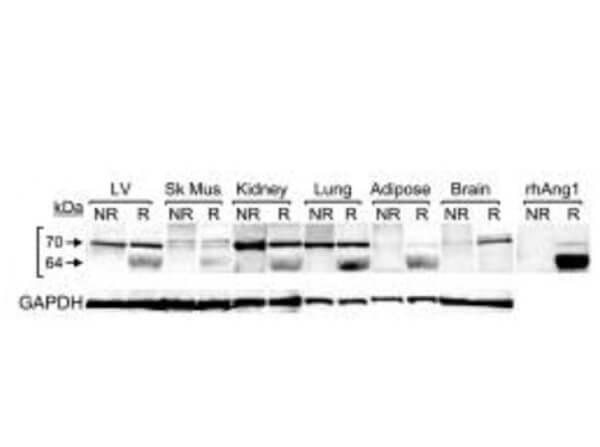
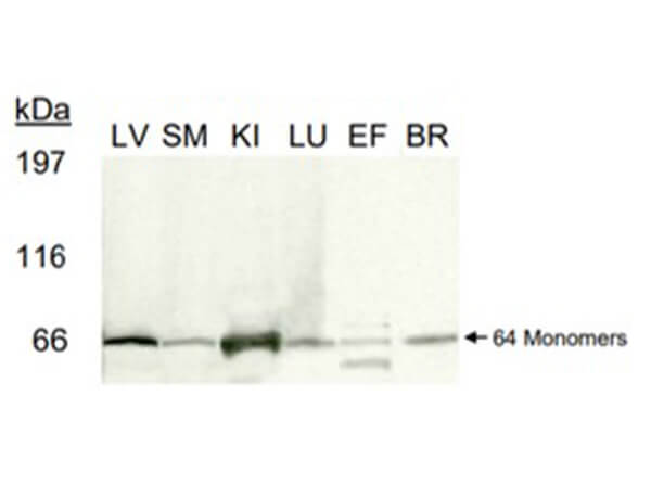
Western blot of Rb Anti-Angiopoietin 1 Antibody. 100µg of protein lysates treated with PNGase F and evaluated under reduced conditions from Human Left Ventricle, skeletal muscle, kidney, lung, epididymal fat tissue, and brain tissue. Anti-Angiopoietin1 incubated and reprobed with Anti-GAPDH antibodies. Dallabrida SM et al. 2008.
Western blot of Rabbit Anti-Angiopoietin 1 Antibody. Tested on 100µg of PNGase-F-treated and reduced lysates: Mouse Left Ventricle, Skeletal Muscle, Kidney, Lung, Epididymal Fat Tissue, and Brain on a 3-8% gel. Anti-Angiopoietin 1 incubated for 1 hr, anti-Rabbit IgG HRP secondary 1:1500 incubated for 1hr. Dallabrida SM et al. 2008.
* Mehrwertsteuer und Versandkosten nicht enthalten. Irrtümer und Preisänderungen vorbehalten